
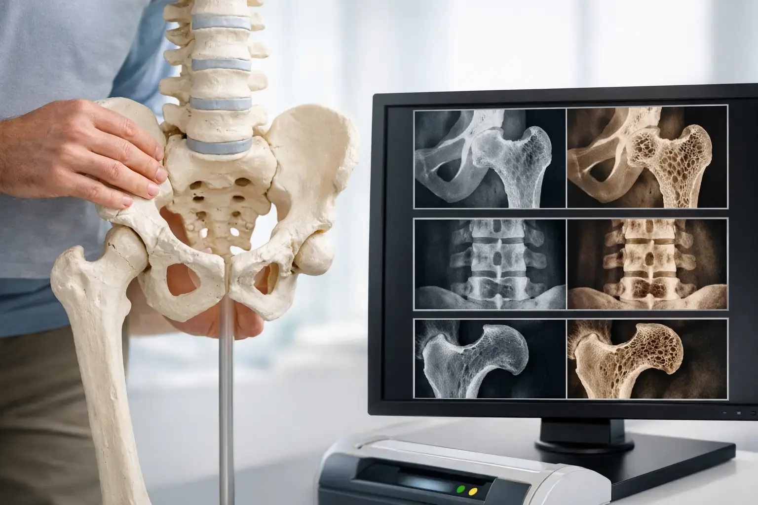
A normal bone-density result can feel like the end of the conversation. For some people - especially those with type 2 diabetes, a history of rapid weight loss, or new concerns during menopause - it may only be the beginning. Can normal density hide fragility? Yes. Density measures an essential part of bone health, but it does not fully describe the signals associated with skeletal fragility or the likelihood of fracture.
That distinction matters because fractures are rarely the first biological change. They are the event that makes an invisible process impossible to ignore. Meaningful bone information should be available before that point.
Can Normal Density Hide Fragility?
Bone mineral density, commonly reported as a T-score or Z-score, estimates how much mineralized bone is present at a measured site. It is useful information and remains central to osteoporosis assessment. Low density is clearly associated with higher fracture risk.
But bone strength is not determined by mineral amount alone. Two people can have similar density measurements and different fracture experiences. One may have a low-density result that correctly signals elevated concern. Another may have density in the normal or osteopenic range, yet show other signs associated with greater fragility.
This is not a contradiction. It reflects the difference between measuring quantity and understanding risk.
Think of two homes built with the same amount of lumber. The total wood tells you something important, but it does not answer every question about how the structures will perform under stress. Bone density similarly tells you how much mineralized bone is present. It cannot, by itself, account for every biological feature that contributes to whether a bone is more or less likely to fracture.
A normal density result is therefore reassuring within its proper meaning: it indicates that the measured mineral content is not low by the relevant reference standard. It is not a complete declaration that fracture risk is absent.
Why Type 2 Diabetes Is the Clearest Example
Type 2 diabetes makes the density-fragility gap especially visible. People with type 2 diabetes may have bone mineral density that appears normal or even higher than expected, while their fracture risk remains elevated in research populations.
One proposed mechanism involves advanced glycation end products, or AGEs. With chronic elevated glucose exposure, these compounds can accumulate in collagen, a protein that is fundamental to bone tissue. Bone mineral is not the whole structure. Collagen is part of the framework in which mineral is organized. Changes in that tissue environment may help explain why density does not always track fracture risk in diabetes as closely as expected.
The practical point is not that a density scan is wrong. It is that density alone can tell an incomplete story in certain conditions.
Type 2 diabetes is not the only situation where this question arises. Long-term glucocorticoid exposure, some cancer-treatment histories, major changes in body weight, and the menopause transition can all prompt a broader discussion of skeletal health. The details differ from person to person. What they share is a reasonable question: beyond how much mineralized bone is present, is there more to understand about fragility and fracture risk?
What Density Can Tell You - and What It Cannot
A density measurement provides a standardized, trackable number. It can identify low bone mass and osteoporosis by established thresholds, help establish a baseline, and show change over time when measurements are performed and interpreted appropriately.
It does not directly provide an independent measure of the reflected ultrasound characteristics associated with fragility. Nor does a density number, standing alone, provide the same kind of integrated estimate of near-term fracture risk that combines density with additional fragility information.
That boundary is worth stating plainly. No single test captures every contributor to fracture. Age, prior fracture, falls, medications, chronic conditions, family history, body weight, and other clinical factors can all matter. A bone scan is one part of a larger clinical picture.
Still, when people are given only density, a meaningful layer of bone information can remain unseen. That is particularly frustrating for someone who is already monitoring glucose, cholesterol, strength, sleep, body composition, or cardiovascular markers. Bones deserve more than a single data point when better information is available.
How REMS Adds Information Beyond Density
REMS, or Radiofrequency Echographic Multi Spectrometry, uses radiation-free ultrasound to assess bone mineral density at the lumbar spine and proximal femur. Those are clinically meaningful sites for bone assessment and fracture risk.
The technology does more than produce a density measurement. It analyzes raw ultrasound signals reflected from bone and derives an independent Fragility Score. The Fragility Score reflects signal features associated with skeletal fragility that density alone does not capture.
That distinction is deliberate. The Fragility Score is not simply another way of restating a T-score. It is separate information derived from the ultrasound signal. REMS then considers density and fragility information together to estimate five-year fracture risk.
A REMS report can therefore answer three connected questions:
- How much mineralized bone is present?
- Is the reflected ultrasound signal showing signs associated with greater fragility?
- What do those measurements indicate together about estimated fracture risk over the next five years?
The point is not to replace clinical judgment with a number. It is to give you and your clinician a more complete starting point for that judgment.
Why Precision Changes the Monitoring Conversation
Monitoring bone health is challenging when the expected biological change is small and the measurement itself has meaningful variability. If a reported change is within the instrument's normal measurement error, it can be difficult to know whether the bone truly changed or the test simply varied.
REMS has an intra-operator precision error below 0.5%, compared with the 0.9% to 1.9% typically reported for DXA at the spine. That difference is not merely technical language. Tighter precision improves the ability to distinguish a meaningful change from measurement noise.
This can matter when a person wants a baseline before a major health transition, is following bone health after an osteoporosis diagnosis, or is trying to understand whether changes over months are measurable. It does not mean every small movement in a report carries clinical significance. It means the measurement is designed to make follow-up information more dependable.
Because REMS uses ultrasound rather than ionizing radiation, repeat measurement does not add radiation exposure. That feature supports a different kind of conversation about bone monitoring: one based on observing change when it may be relevant, rather than waiting years simply because repeat imaging carries a radiation burden.
A More Useful Question Than “Is My Density Normal?”
“Is my density normal?” is a fair question. A better one may be: “What does my density tell me, and what does it leave unanswered?”
For someone with type 2 diabetes, a normal density result may deserve context rather than dismissal. For a person in perimenopause, a baseline can make future change visible. For someone already told they have osteopenia or osteoporosis, an independent Fragility Score can add information without minimizing what the density result already shows.
The goal is not to turn every normal result into a source of concern. Most bone-health decisions do not rest on one number, and they should not rest on fear. The goal is to avoid confusing a partial answer with a complete one.
MobileREMS provides density, an independent Fragility Score, and an estimated five-year fracture risk in one radiation-free assessment. The resulting report is information you can understand, follow over time, and bring into a conversation with your physician or care team.
Your bones are changing tissue, not a static score. Asking for information that reflects both bone quantity and signs associated with fragility is a practical way to make the invisible more understandable - before a fracture is asked to explain it for you.