
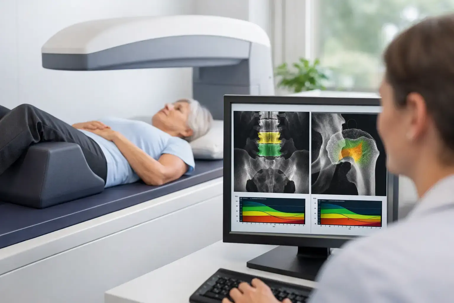
A fracture is often the first event that makes bone loss visible. By then, the underlying process may have been unfolding quietly for years. Osteoporosis screening changes that sequence by making bone health measurable before a fall, a broken wrist, a vertebral compression fracture, or a hip fracture forces the conversation.
The question is not only whether you qualify for a scan under a guideline. It is also what information the scan can give you. Bone mineral density remains central to skeletal assessment, but density alone does not fully explain why two people with similar T-scores can have different fracture experiences.
What osteoporosis screening is designed to find
Osteoporosis is a condition of reduced bone strength that raises fracture risk. Bone strength is influenced by the amount of mineralized bone present, along with qualities of the bone associated with fragility. A screening assessment aims to identify low bone density and clarify fracture risk before a low-trauma fracture occurs.
The most familiar measurement is bone mineral density, usually reported as a T-score. The T-score compares your density with the average peak bone density of a healthy young adult of the same sex. A T-score of -2.5 or lower meets the diagnostic threshold for osteoporosis. A score between -1.0 and -2.5 is commonly described as osteopenia, or low bone mass.
Those categories are useful, but they are not the whole clinical picture. A T-score tells you how much mineralized bone is present at the site measured. It does not, by itself, tell you everything about the structural signs associated with fragility or your near-term likelihood of fracture.
That distinction matters because bones do not all behave the same way at the same density. Think of two people whose scans show similar bone mineral density at the hip. One may have ultrasound signal characteristics associated with greater fragility, while the other does not. The density number is similar; the information about fragility is not.
Who may benefit from earlier bone information
Age-based recommendations have an essential role in public health. In the United States, routine screening is commonly recommended for women beginning at age 65, and for younger postmenopausal women whose risk is elevated. Yet eligibility rules are not a biological start date for bone change.
For many women, the menopause transition is a meaningful window. Falling estrogen can accelerate bone loss, while routine screening may still be years away. An earlier baseline can show where density stands and make future change easier to interpret.
Risk is also not limited to menopause or age. A history of low-trauma fracture, a parent with a hip fracture, smoking, heavy alcohol exposure, low body weight, prolonged immobility, and certain medical conditions or medications can all shift the risk conversation. Long-term glucocorticoid use is a well-recognized example because these medications can affect bone formation and resorption.
Type 2 diabetes deserves particular attention. It can produce a false sense of reassurance: bone density may be normal or even higher than expected, while fracture risk remains elevated. The mechanism is complex and likely includes changes in bone material properties associated with chronic high glucose, falls risk, and diabetes-related complications. The practical point is clear: a normal density result does not always mean normal skeletal risk.
Cancer-treatment histories, rapid weight loss, and the use of GLP-1 medications can also bring bone health into focus. The relevant question varies by person and context, but the need for objective baseline information is often the same. A scan can give you and your clinician a starting point for a more informed discussion.
What a standard density scan can and cannot answer
DXA, or dual-energy X-ray absorptiometry, has long been the standard method for measuring bone mineral density. It is a validated, widely used tool for diagnosis and monitoring. Its value should not be minimized.
But a density score answers one question: how much mineralized bone is present at the measured site? It cannot independently characterize every contributor to fragility. This is one reason fracture risk assessment considers more than T-score, including age, prior fractures, medical history, medication exposure, and other clinical factors.
A useful scan should not replace clinical judgment. It should make clinical judgment better informed. If you already have osteopenia or osteoporosis, density remains necessary information. If you are deciding whether to begin, change, or monitor a bone-health strategy, knowing density is still part of the picture. The limitation is treating that one number as the entire picture.
What REMS adds to osteoporosis screening
Radiofrequency Echographic Multi Spectrometry, or REMS, uses radiation-free ultrasound to measure bone mineral density at the lumbar spine and proximal femur. Those are the same clinically meaningful regions commonly assessed in bone-density testing.
REMS also analyzes the raw ultrasound signals reflected from the bone. From those signals, it derives an independent Fragility Score, reflecting signal patterns associated with skeletal fragility that density alone does not capture. It then weighs density and the Fragility Score together to estimate five-year fracture risk.
This produces three pieces of information that can be followed over time: bone mineral density, Fragility Score, and estimated five-year fracture risk. The Fragility Score is not a substitute for density, nor does it diagnose every aspect of bone quality. It is additional information, derived through a different part of the measurement, that helps clarify the gap between how much bone is present and how fragile that bone may appear.
For someone with type 2 diabetes, this can be especially relevant. A density result may look reassuring while the broader fracture-risk story is less simple. For someone entering menopause, the value may be a baseline that includes more than density. For someone already living with osteopenia, it may offer another way to understand whether low density and fragility are telling the same story.
MobileREMS uses FDA-cleared Echolight REMS technology. Because the test is radiation-free and has reported intra-operator precision error below 0.5%, it can support repeat measurement over shorter intervals when clinically appropriate. Precision is not a technical detail. If a measurement changes, lower error helps distinguish a meaningful biological shift from ordinary measurement noise.
Preparing for a useful bone-health conversation
The most valuable result is one that can be put in context. Bring prior bone-density reports if you have them, along with information about fractures, family history, medication exposure, menstrual or menopause history, diabetes, cancer treatment, and major weight changes. These details do not change the scan itself, but they can change how its findings are interpreted.
It also helps to know what a result cannot settle on its own. No single scan can predict exactly whether you will fracture. Fractures arise from both skeletal vulnerability and the circumstances of a fall. A result may clarify risk without dictating a single next step, and an apparently reassuring result does not erase every relevant risk factor.
Bone is living tissue. Mechanical loading stimulates bone adaptation, nutrition and vitamin D contribute to bone metabolism, hormones influence turnover, and some diseases and treatments alter that balance. Screening does not turn those realities into a simple score. It gives you a clearer view of where you are within them.
The goal is not to become afraid of an invisible process. It is to make that process visible enough to discuss, monitor, and understand before consequences have to do the explaining.