
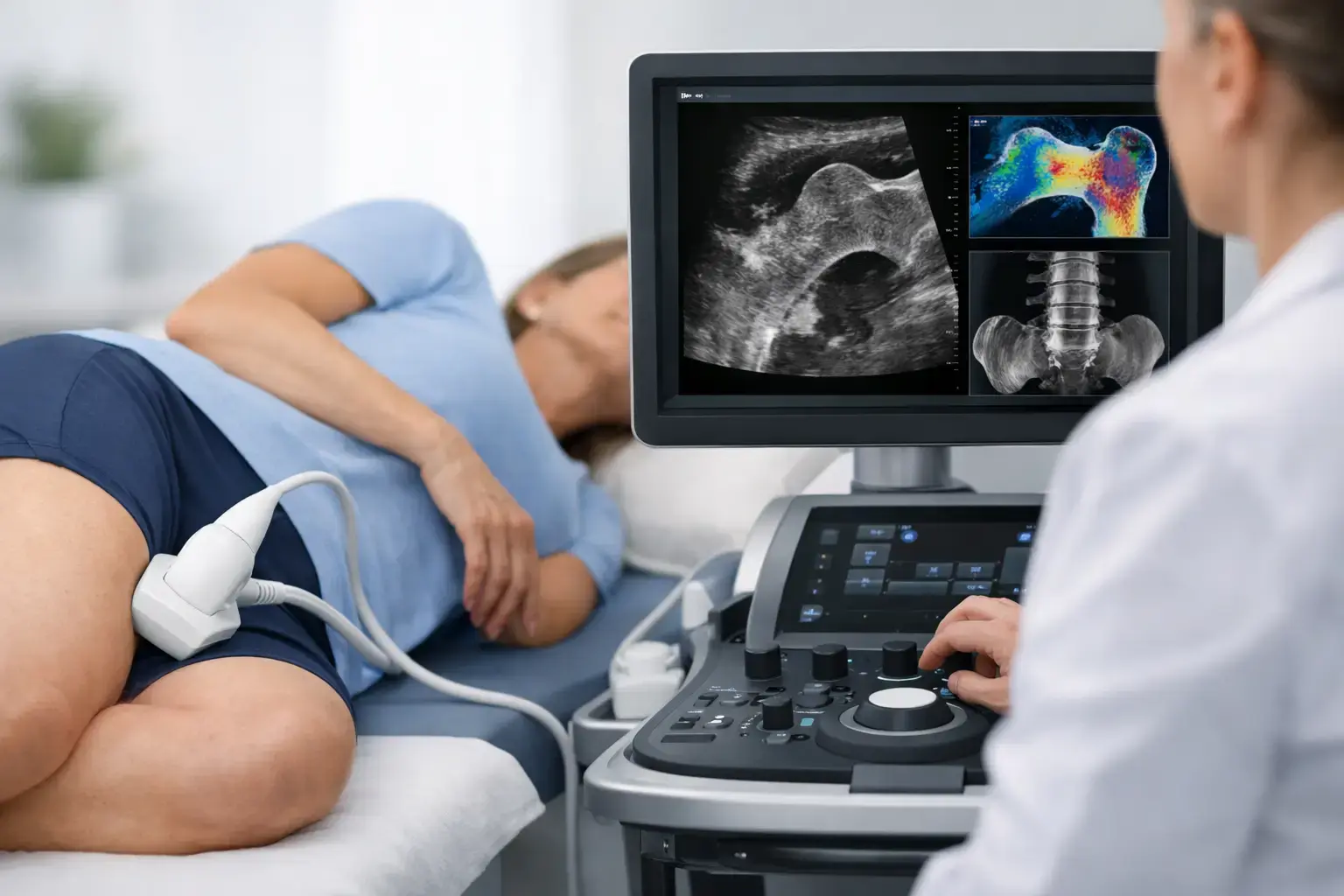
A normal-looking density result can be reassuring, but it may not be the whole story. Ultrasound bone density test accuracy depends on what kind of ultrasound is used, where it measures, and what the result is designed to tell you about your bones.
That distinction matters most when the question is larger than, “How much mineralized bone do I have?” If you are in menopause, have type 2 diabetes, are monitoring bone health during rapid weight loss, or simply want a baseline before a consequence forces the issue, you may also want to know whether your bone is showing signals associated with greater fragility and what that means for fracture risk.
Accuracy is not one number
When people ask whether an ultrasound bone density test is accurate, they are often asking several different questions at once. Does it measure bone mineral density reliably? Does it agree with established density testing? Can it detect a meaningful change over time? Does it help identify people at greater fracture risk?
Those are related questions, but they are not interchangeable.
A test can be highly precise, meaning repeated measurements are very close together, without answering every question about fracture risk. A test can also correlate well with another density test while providing no additional information beyond that test. The most useful assessment depends on whether you need a screening signal, a density measurement at clinically meaningful skeletal sites, a way to monitor change, or a broader view of risk.
For bone health, accuracy has to be tied to purpose.
Not all bone ultrasounds measure the same thing
Many people first encounter bone ultrasound as a heel screening test. These devices, often called quantitative ultrasound or QUS, assess the calcaneus, the heel bone. They may use ultrasound transmission characteristics such as speed of sound and broadband ultrasound attenuation to help identify people who may benefit from further evaluation.
That can be useful as a screening approach. But a heel measurement is not the same as measuring bone mineral density at the lumbar spine and proximal femur, the sites commonly used in osteoporosis assessment and where many clinically significant fractures occur.
REMS, or radiofrequency echographic multi-spectrometry, is a different ultrasound-based technology. It measures bone mineral density at the lumbar spine and proximal femur using ultrasound imaging. It then analyzes the raw radiofrequency signals reflected from bone rather than relying only on the final image.
That difference in site and signal analysis is central to what REMS can report. A REMS scan provides density, but it also derives an independent Fragility Score from reflected ultrasound signals and combines the two to estimate five-year fracture risk.
What REMS accuracy means in practice
REMS is FDA-cleared for bone mineral density assessment at the lumbar spine and proximal femur. Its bone mineral density measurements have been clinically validated against DXA, the established reference method for diagnosing osteoporosis based on BMD.
Still, “agrees with DXA” is only part of the accuracy conversation. DXA measures are affected by factors that can become more common with age, including degenerative changes in the spine, vascular calcification, and positioning differences. No test is interpreted well by treating every number as self-explanatory. The scan site, image quality, clinical context, and previous measurements all matter.
One of REMS's practical strengths is precision. MobileREMS uses technology with an intra-operator precision error below 0.5%. By comparison, precision errors commonly reported for DXA at the spine are roughly 0.9% to 1.9%.
That may sound like a small technical difference. It is not. Precision determines how confidently a clinician can separate a real biological change from measurement noise. If a method varies by 1% simply because of the measurement process, a small change on a later report may be difficult to interpret. Lower measurement error makes earlier re-measurement more meaningful, including when someone is tracking bone health through a period when change may be occurring faster.
Precision is not the same as accuracy, but both matter. A meaningful bone-health measurement needs to be reliable enough to repeat and clinically grounded enough to interpret.
Density and fragility are related, not identical
Bone mineral density tells you how much mineralized bone is present in the area measured. It is a valuable number and remains foundational to bone assessment. But density alone does not fully explain why some people fracture and others do not.
Consider two people with similar density results. One may have a lower Fragility Score and a lower estimated fracture risk. The other may show ultrasound signal characteristics associated with greater skeletal fragility, resulting in a higher Fragility Score and different five-year fracture-risk estimate. Their density may look similar, while the information beyond density does not.
REMS does not claim to measure every property that affects whether a bone fractures. Fracture risk is also influenced by age, prior fracture, falls, medications, medical conditions, and other factors. It does, however, add an independent signal related to fragility that density alone does not capture.
This is especially relevant in conditions where the density story can look falsely reassuring.
Type 2 diabetes is the clearest example
People with type 2 diabetes can have bone mineral density that reads as normal or even elevated while their fracture risk remains higher than density alone would suggest. Research has repeatedly documented this mismatch. The reasons are complex and may include changes related to long-term glucose exposure and bone remodeling, along with diabetes-related fall risk and complications.
The point is not that a density result is wrong. It is that density is answering one question. When the question is fracture risk, a normal density result does not always close the conversation.
The same principle can matter with long-term glucocorticoid exposure, cancer-treatment histories, substantial weight loss, and other situations that affect skeletal health. Your health history gives the result context. The result, in turn, gives your physician more objective information to consider.
How the ultrasound bone density test is performed
A REMS examination is radiation-free and noninvasive. During the scan, an ultrasound probe is used to assess the lumbar spine and proximal femur. The system identifies the relevant bone region and analyzes reflected raw ultrasound signals to calculate BMD and the Fragility Score.
The experience is different from a whole-body imaging study. There is no ionizing radiation and no enclosed scanner. You leave with a report that includes three understandable measures: bone mineral density, Fragility Score, and estimated five-year fracture risk.
That report is designed to support a clinical conversation, not replace one. A scan cannot determine why a change has occurred, whether a medication is appropriate, or what an individual treatment plan should be. It can make the invisible more visible: how much mineralized bone is present, whether the signals show greater fragility, and what those measures suggest together about risk.
When an ultrasound result is most useful
The value of a bone assessment is often highest before a fracture, not after one. That does not mean everyone needs the same test at the same time. It means that a person with a reason to ask should not have to settle for less information than the question requires.
For someone entering menopause, a baseline can establish where bone health begins before years of change accumulate. For someone with osteopenia or osteoporosis, repeatable measurements may help make follow-up conversations more specific. For the person who tracks lipids, glucose, muscle mass, and cardiovascular fitness but has never measured their bones, the gap is worth noticing: peak bone mass is largely built by early adulthood, and bone loss can begin long before symptoms appear.
A fracture is often the first visible event in a process that has been silent for years. Better information does not predict every outcome, but it gives you and your care team a clearer starting point for the decisions ahead.