
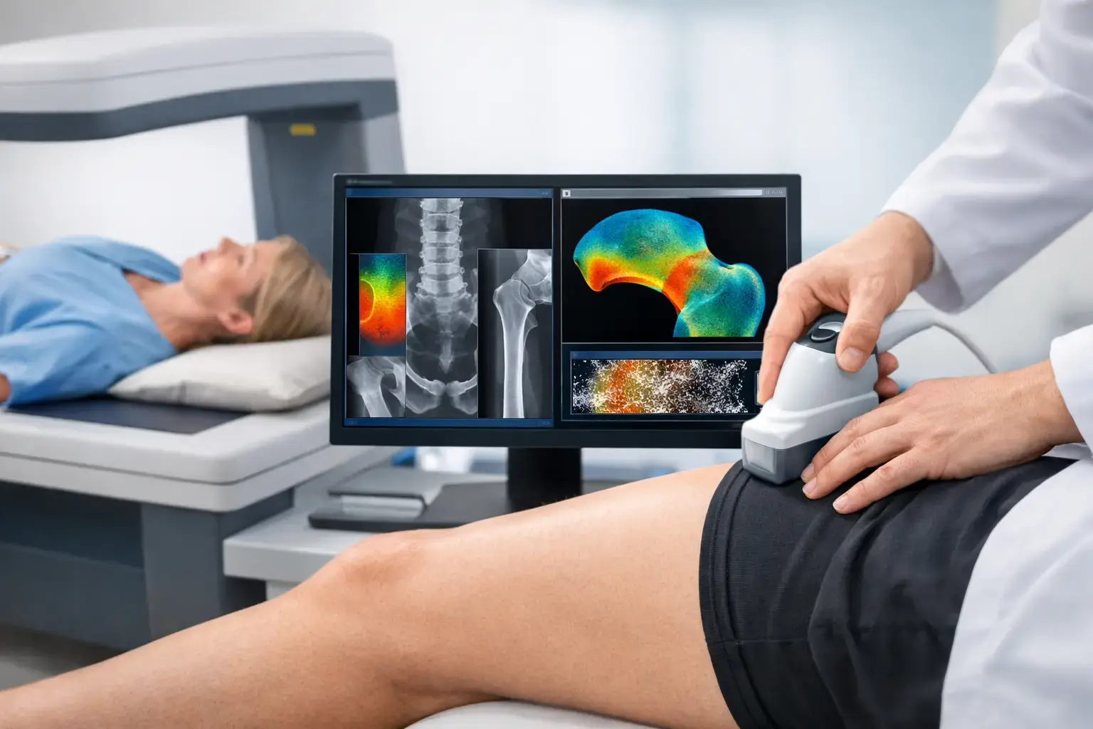
A DXA vs REMS bone scan comparison matters most when a single T-score does not feel like the whole answer. Maybe you are entering menopause, living with type 2 diabetes, monitoring bone changes during weight loss, or simply wondering why you track cholesterol and glucose more closely than the structure that carries you through every day.
Both tests can measure bone mineral density. But they do not ask precisely the same question of your skeleton. DXA measures how much mineralized bone is present. REMS measures density too, then analyzes reflected ultrasound signals to derive an independent Fragility Score and estimate five-year fracture risk.
That distinction is not semantic. Two people can have similar density results and different signs of skeletal fragility. Density remains valuable information. It is just not the entire picture.
DXA vs REMS bone scan: the central difference
DXA, short for dual-energy X-ray absorptiometry, has been the established standard for measuring bone mineral density for decades. It uses a very low dose of X-ray radiation to estimate the mineral content of bone, commonly at the lumbar spine and hip. The report typically includes a T-score, which compares your density with that of a healthy young adult reference population, and sometimes a Z-score, which compares it with an age-matched population.
Those numbers serve an important clinical role. DXA is widely used in osteoporosis diagnosis, treatment decisions, and longitudinal care. It is also deeply embedded in clinical guidelines, insurance processes, and physician workflows. If your clinician has been following your DXA results over time, those prior measurements remain meaningful context.
REMS, or radiofrequency echographic multi-spectrometry, takes a different measurement route. It uses ultrasound rather than ionizing radiation to measure bone mineral density at the lumbar spine and proximal femur. The technology then analyzes the raw radiofrequency signals reflected from the bone. From those signals, it derives a Fragility Score that is independent of density.
The result is not a claim that REMS sees every property that affects whether a bone fractures. It does not directly measure bone strength, flexibility, microarchitecture, or failure load. What it does provide is an additional signal associated with fragility, alongside density. REMS then weighs density and Fragility Score together to estimate fracture risk over the next five years.
Put simply, DXA answers, “How much mineralized bone is there?” REMS answers that question and adds, “Is this bone showing ultrasound-signal characteristics associated with greater fragility?”
Why density can miss part of the risk story
Bone mineral density is strongly associated with fracture risk, which is why it has long been central to bone-health assessment. Yet fracture risk is not determined by density alone. Age, prior fractures, falls, medications, health conditions, and the biological quality of bone all shape the picture.
Type 2 diabetes is a clear example of why a reassuring density result can be incomplete. People with type 2 diabetes may have normal or even higher bone mineral density while still facing elevated fracture risk. Researchers believe altered bone material properties, including changes related to chronic high blood glucose and glycation, may contribute to this mismatch. A density number can look favorable while the fracture-risk question remains more complicated.
Imagine two people whose density falls in the same range. A DXA report may make them appear similar on that one measure. If their REMS Fragility Scores differ, however, the REMS assessment may indicate that the underlying fragility signals are not the same. That does not make one result “right” and the other “wrong.” It shows why density and fragility should not be treated as interchangeable concepts.
This gap can also matter for people using long-term glucocorticoids, those with a history of cancer treatment, and people experiencing substantial or rapid weight loss. These circumstances do not guarantee bone problems. They do make it reasonable to seek information that is broader than density alone.
Radiation, comfort, and what the scan experience is like
The radiation difference between the two tests is straightforward. DXA uses a low dose of X-ray radiation. REMS uses ultrasound and is radiation-free.
For many people, the low radiation dose from DXA is not a practical barrier. DXA has a long safety record and remains an appropriate test in many settings. But radiation-free measurement may matter if you value repeat assessment without radiation exposure, are building an earlier baseline, or want a monitoring approach that can be used more frequently when clinically relevant.
A REMS scan is performed with an ultrasound probe over the lumbar spine and hip area. It does not involve an injection, contrast material, or compression. As with any imaging test, positioning and trained operation matter because measurement quality depends on obtaining and interpreting a reliable signal.
Neither exam is a general health check or a substitute for a clinician’s evaluation. A scan result becomes more useful when it is considered alongside your health history, medications, fracture history, and the reason you are assessing your bones in the first place.
Precision changes what monitoring can mean
One of the most practical differences is precision: how consistently a device can reproduce a measurement when your bones themselves have not changed.
Small shifts in a bone-density report can be difficult to interpret if the instrument’s normal measurement variation is close to the size of the observed change. This is why interval monitoring with DXA is often spaced over longer periods. A result needs to move enough to distinguish biological change from measurement noise.
Echolight REMS technology has reported an intra-operator precision error below 0.5%, compared with approximately 0.9% to 1.9% commonly reported for DXA at the spine. Precision figures vary by site, device, operator, and study design, so no single percentage answers every real-world question. Still, lower measurement error has a meaningful implication: it can make smaller changes more interpretable.
That matters when skeletal change may be occurring over months rather than years. A repeat measurement is only helpful if a difference on the report is likely to reflect a difference in bone, not ordinary noise from the test.
When DXA remains the right choice
DXA is not obsolete, and REMS is not a reason to discard decades of DXA data. DXA may be the most practical option when your care team uses it for diagnostic classification, when prior DXA results are the key comparison point, or when a health system and insurer require DXA within an established workflow.
DXA also has a particularly familiar language in medicine. Many clinicians can immediately place a T-score in the context of guidelines and prior records. That continuity has value.
At the same time, a familiar test is not automatically a complete test for every question. If you want density plus an independent fragility measure and a five-year fracture-risk estimate, REMS provides information that DXA alone does not generate. The strongest choice depends on what information is missing, not on declaring one modality the winner in every situation.
When REMS adds the most
REMS is especially relevant when you want a baseline before routine screening would typically occur, or when a density-only answer feels insufficient. That may include perimenopause, a new osteopenia or osteoporosis diagnosis, type 2 diabetes, medication-related risk, or preparation for a discussion with a physician or surgical team.
It can also fit a prevention-minded approach. Peak bone mass is largely built by the late twenties, genetics influences much of the ceiling, and bone loss can begin well before many people ever receive a routine scan. By midlife, many people have detailed data on body composition, metabolic markers, and cardiovascular risk but no objective picture of their bone density or fragility.
A REMS report provides three trackable measures: bone mineral density, Fragility Score, and estimated five-year fracture risk. Those numbers do not predict an individual future with certainty. They can make a conversation more specific - whether that conversation is about a changing life stage, an existing diagnosis, or how to monitor a known risk factor over time.
The better question is not “Which scan is best?”
The useful question is: what do you need to know about your bones that you do not know yet?
If you need an established density measurement integrated into conventional diagnostic pathways, DXA is often central to the answer. If you want density alongside information about fragility signals and a five-year risk estimate, REMS expands the conversation. For some people, both can be useful at different points in care.
Bone health should not become visible only after a fracture forces attention. A clear baseline and the right kind of follow-up can give you something more useful than reassurance or alarm: a better understanding of what your bones are telling you now.