
Most adults can tell you their cholesterol number or last blood pressure reading. Far fewer know what is happening in their skeleton until a fall, a fracture, or an unexpected scan creates the question. When should adults check bones? Earlier than many people assume when a meaningful risk factor, life transition, or treatment decision is already in view.
Bone loss is usually silent. You do not feel mineralized bone declining, and a bone does not announce that it is becoming more fragile. By the time a low-trauma fracture occurs, the biology has often been changing for years. The purpose of checking bone health is not to predict disaster. It is to replace a blind spot with useful information before consequences make the conversation urgent.
When should adults check bones?
There is no single age that fits every adult. Formal screening guidelines are built to identify populations most likely to benefit from testing, while an individual assessment also depends on menopause status, medical history, medications, prior fractures, and decisions you are facing now.
For many women, routine bone-density screening begins at age 65. The U.S. Preventive Services Task Force also supports screening for postmenopausal women younger than 65 who have increased osteoporosis risk. Organizations such as the Bone Health and Osteoporosis Foundation commonly identify age 70 as a routine screening point for men, with earlier assessment for men ages 50 to 69 who have risk factors. Evidence for population-wide screening recommendations in men remains less settled, which is different from saying bone health is not relevant to men.
Those age thresholds are useful public-health guardrails, not a biological starting gun. Several situations can make a baseline bone assessment worth discussing well before routine screening age.
1. The menopause transition
Estrogen helps regulate bone remodeling, the continuous cycle in which older bone is broken down and new bone is formed. During perimenopause and menopause, that balance can shift toward faster loss. The pace varies widely, which is exactly why age alone is an incomplete guide.
A baseline during this transition can establish where you are starting from rather than leaving you to infer years later what may have changed. It can be particularly useful if you have a family history of hip fracture or osteoporosis, early menopause, low body weight, a previous fracture, or conditions that affect nutrient absorption or hormone regulation.
2. After a fracture that seemed out of proportion to the fall
A fracture after a fall from standing height or less deserves a bone-health conversation, especially after age 50. Not every fracture has the same implication. A finger broken during a high-impact sports injury is different from a wrist fracture after a routine stumble. Context matters.
Still, a fracture can be the first visible outcome of an invisible process. Assessment helps clarify whether low bone mineral density may be part of the picture and whether other information about skeletal fragility changes how that density result should be understood.
3. When type 2 diabetes is part of your health history
Type 2 diabetes is one of the clearest examples of why density cannot answer every bone question alone. Research has repeatedly found that people with type 2 diabetes can have bone mineral density that appears normal, or even higher than expected, while their fracture risk remains elevated.
The mechanism is still being studied, but long-term high blood glucose can alter bone material through glycation-related changes, and diabetes-related complications may also increase fall risk. The practical point is direct: a reassuring density number does not always tell the full fracture-risk story. If you have type 2 diabetes, asking only, “How much bone do I have?” may leave out the more consequential question: “Is there evidence that this bone is more fragile than its density suggests?”
4. During medications or treatments that affect bone
Long-term glucocorticoid use is a well-established concern because these medications can disrupt bone remodeling and reduce bone formation. Certain cancer treatments can affect sex hormones and bone turnover. Conditions involving inflammatory disease, celiac disease, kidney disease, thyroid disorders, or reduced nutrient absorption can also change the bone-health conversation.
The appropriate timing depends on the condition, the treatment, and the clinical decision at hand. For someone beginning a therapy with known skeletal effects, a baseline may be useful because it creates a reference point. For someone already in treatment, the conversation is usually less about checking a box and more about understanding whether meaningful change is occurring over time.
5. During rapid weight loss or major body-composition change
Weight change can be metabolically beneficial while still raising legitimate questions about the skeleton. Bone responds to loading, hormones, nutrition, and energy availability. When body weight changes rapidly, those signals may change too.
This does not mean that intentional weight loss is inherently harmful to bone, nor does it mean every person using a GLP-1 medication needs the same testing schedule. It means the skeleton belongs in the conversation, particularly when weight loss is substantial, nutritional intake has changed, or other bone-risk factors are present. A baseline provides context that cannot be recreated later.
6. Before a decision where bone information matters
Sometimes the right time is not tied to an age or diagnosis. It is tied to a decision. You may be considering osteoporosis medication, preparing for an orthopedic procedure, reviewing a family history, or building a prevention-focused health plan. Objective information can make that conversation more specific.
This is especially relevant for adults who track cardiovascular markers, glucose, fitness, and body composition but have never measured their bones. Peak bone mass is largely built by early adulthood, with genetics influencing much of the ceiling. From midlife onward, the relevant question is often not whether you can rebuild the skeleton you might have had at 25. It is whether you understand your present baseline and can observe the direction of change.
Why bone density is necessary but not always sufficient
A standard bone-density result estimates how much mineralized bone is present at a measured site, commonly reported as a T-score or Z-score. It is valuable information. Lower density is associated with higher fracture risk, and density testing remains central to osteoporosis assessment.
But density is not synonymous with fragility. Consider two people with similar bone mineral density at the hip. One may have ultrasound signal characteristics associated with greater skeletal fragility, while the other does not. If you look only at density, they appear to tell the same story. They may not carry the same estimated fracture risk.
That distinction matters most when the clinical picture and the density result do not neatly agree: the person with type 2 diabetes and a normal density score, the person with osteopenia who wants more clarity about risk, or the person monitoring changes during a period when bone may be changing quickly.
What REMS adds to a bone-health assessment
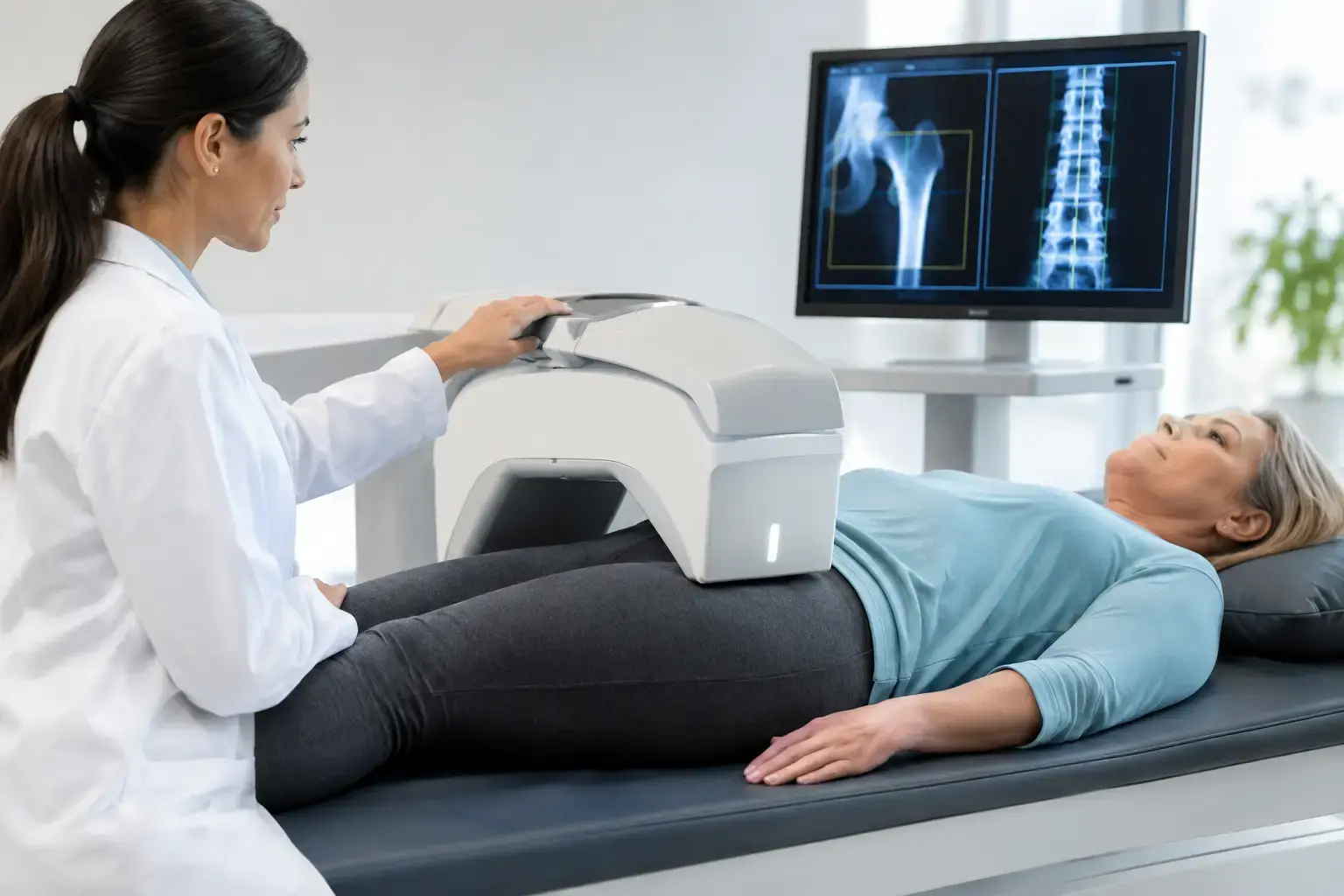
Radiofrequency Echographic Multi Spectrometry, or REMS, uses radiation-free ultrasound to measure bone mineral density at the lumbar spine and proximal femur. It also analyzes the raw ultrasound signals reflected from bone to derive an independent Fragility Score.
The Fragility Score is not another name for density. It reflects signal features associated with skeletal fragility that density alone does not capture. REMS then considers density and Fragility Score together to estimate five-year fracture risk.
That produces three understandable pieces of information: how much mineralized bone is present, whether the reflected signal shows signs associated with greater fragility, and what the combination may mean for fracture risk over the next five years. It does not replace a clinician's interpretation of your full history, fall risk, medications, or other health conditions. It adds a different layer of objective information.
Precision also changes what monitoring can mean. REMS has an intra-operator precision error below 0.5%, compared with commonly reported DXA spine precision ranges of roughly 0.9% to 1.9%. Measurement error is not just a technical detail. If the expected change is small, you need confidence that a difference on a report is more than normal instrument variation. Tighter precision can support meaningful reassessment over shorter intervals when a clinician and patient have a reason to monitor.
The useful question is not “Am I old enough?”
Age-based screening remains important, and adults who meet established screening criteria have a clear reason to act. But a more useful question is whether your present circumstances create a reason to understand your bones now.
If menopause, diabetes, a prior fracture, medication exposure, rapid weight loss, or an upcoming care decision has put bone health on your mind, that is already meaningful information. A bone assessment cannot erase uncertainty, but it can turn a vague concern into a clearer conversation with your clinician - before a fracture has to start it for you.