
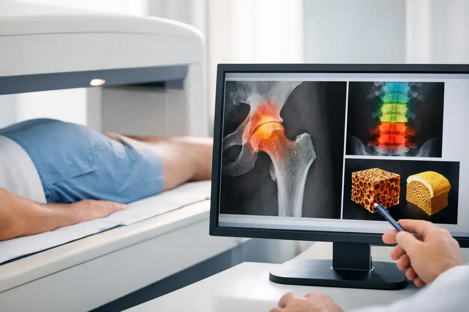
A normal bone density result can be falsely reassuring when you have type 2 diabetes. Diabetes bone tests deserve a closer look because people with type 2 diabetes may have bone mineral density that reads as normal, or even higher than expected, while their fracture risk remains elevated.
That apparent contradiction is not a testing error. It reflects a limitation of treating density as the full story of bone strength. Density tells you how much mineralized bone is present. A fracture happens when a real bone, in a real body, encounters force it cannot withstand. Diabetes can affect that equation in ways a density number does not fully capture.
Why type 2 diabetes can raise fracture risk
The association between type 2 diabetes and fracture has been documented in large population studies, including the Rotterdam Study: higher bone mineral density did not erase the increased fracture risk seen in people with type 2 diabetes. This is the false-normal problem. The scan may show an amount of mineralized bone that appears reassuring, while other diabetes-related changes still matter.
One proposed mechanism involves advanced glycation end products, often called AGEs. With persistent exposure to elevated glucose, sugars can bind to proteins in tissues throughout the body, including collagen. Collagen is a major component of bone's organic matrix. Research suggests glycation can alter the material properties of that matrix and its normal turnover. Bone density testing does not directly measure those changes.
Diabetes may also affect bone remodeling, the continuous process in which older bone is removed and new bone is formed. Peripheral neuropathy, vision changes, reduced muscle function, and episodes of low blood sugar can raise fall risk as well. Those factors do not mean a fracture is inevitable. They do mean that a single density score may not answer the question you actually care about: what does this mean for my likelihood of fracture?
Type 1 diabetes carries its own, often substantial, skeletal risk profile and can be associated with lower density. But type 2 diabetes is the clearest example of why a normal density result should not automatically close the bone-health conversation.
What diabetes bone tests are trying to answer
There is no single test that captures every factor affecting fracture risk. A useful assessment combines different kinds of information, each with a distinct job.
A clinical review starts with your history: diabetes type and duration, prior fractures, falls, medication exposure, kidney disease, menopause status, weight change, smoking, alcohol use, and family history. Long-term glucocorticoid use is particularly relevant because it can affect bone through a different mechanism than diabetes. The purpose is context. A test result has more meaning when it is interpreted alongside the conditions that may influence bone and falls.
A DXA scan is the standard test for bone mineral density, usually measured at the lumbar spine and hip. It provides T-scores and Z-scores, which help clinicians classify low bone mass and osteoporosis and follow density over time. DXA remains useful. The issue is not that density is unhelpful. The issue is that density alone is incomplete for many people with diabetes.
A fracture-risk calculation may also be used. These models combine clinical factors and, often, femoral-neck density to estimate the probability of fracture over a defined period. They are helpful for organizing risk factors, but studies have raised concern that conventional risk calculators can underestimate fracture risk in type 2 diabetes unless diabetes-related risk is specifically considered. A calculated risk is an estimate, not a verdict.
Depending on your clinical picture, a physician may also consider vertebral imaging. Vertebral fractures can occur with little or no memorable injury and may be found incidentally. Identifying an existing vertebral fracture changes the conversation because a prior fragility fracture is one of the strongest indicators of future fracture risk.
Laboratory testing answers a different question again. Tests such as calcium, vitamin D, kidney function, thyroid-related markers, parathyroid hormone, and other studies may help identify contributors to bone loss or altered bone metabolism. An A1C measures average glucose exposure, not bone strength. It belongs in the broader health picture, but it is not a bone test.
Where density leaves an information gap
Imagine two people with similar hip density. On paper, their scans may look nearly identical. One has no major bone-related risk factors. The other has lived with type 2 diabetes for years, has experienced a recent fall, and has a history that suggests more than density alone is contributing to fracture risk.
The density result has not become wrong for the second person. It has become insufficient on its own.
This distinction matters because diabetes-related bone risk is not fully explained by the amount of mineralized bone present. If you only measure quantity, you may miss signals associated with fragility. That is why a normal or high density result in type 2 diabetes should be interpreted with care rather than treated as a blanket reassurance.
The same principle applies beyond diabetes. People using glucocorticoids, undergoing rapid weight loss, taking medications that affect metabolism, or moving through menopause may have reasons to ask for more than a single density number. The appropriate testing pathway depends on the individual and the decision at hand.
REMS adds information beyond bone density
Radiofrequency Echographic Multi Spectrometry, or REMS, measures bone mineral density at the lumbar spine and proximal femur using radiation-free ultrasound. It also analyzes the raw ultrasound signals reflected from bone to derive an independent Fragility Score.
That distinction is central. The Fragility Score is not another way of stating bone density, and it is not a direct measurement of collagen quality, microarchitecture, flexibility, brittleness, or failure load. It reflects ultrasound signal characteristics associated with skeletal fragility that density alone does not capture.
REMS then considers density and the Fragility Score together to estimate five-year fracture risk. Instead of leaving with one number, you leave with three understandable pieces of information: bone mineral density, Fragility Score, and estimated five-year fracture risk.
For someone with type 2 diabetes, this can be particularly useful. A density result may still be normal. The independent fragility information may tell a more complicated story, or it may support the reassuring density result. Either outcome is more informative than assuming density settles the question.
REMS is FDA-cleared and radiation-free. Its reported intra-operator precision error is below 0.5%, compared with the roughly 0.9% to 1.9% commonly reported for DXA at the spine. Precision matters when you are monitoring change. If a method has less measurement noise, smaller shifts can be interpreted with greater confidence, though every result still needs to be considered in clinical context.
Questions worth bringing to your bone-health conversation
If you have diabetes, the most useful question is not simply, “Do I need a bone scan?” It is, “Does my current assessment account for the ways diabetes can affect fracture risk beyond density?”
You might also ask whether your history suggests a need for vertebral imaging, whether your fracture-risk estimate accounts for diabetes, and whether laboratory findings or medications could be contributing to skeletal risk. If you already have a DXA result, ask what it can answer clearly and what it cannot.
For people in Seattle, Kitsap County, Phoenix, and surrounding communities, MobileREMS provides a way to add density, an independent Fragility Score, and a five-year fracture-risk estimate to that discussion without radiation exposure.
Your bones are not a report card on diabetes management, and a normal density result is not a reason to ignore meaningful questions. It is one piece of evidence. The goal of better testing is not to predict disaster. It is to make an invisible part of your health more understandable before a fracture is what finally brings it into view.