
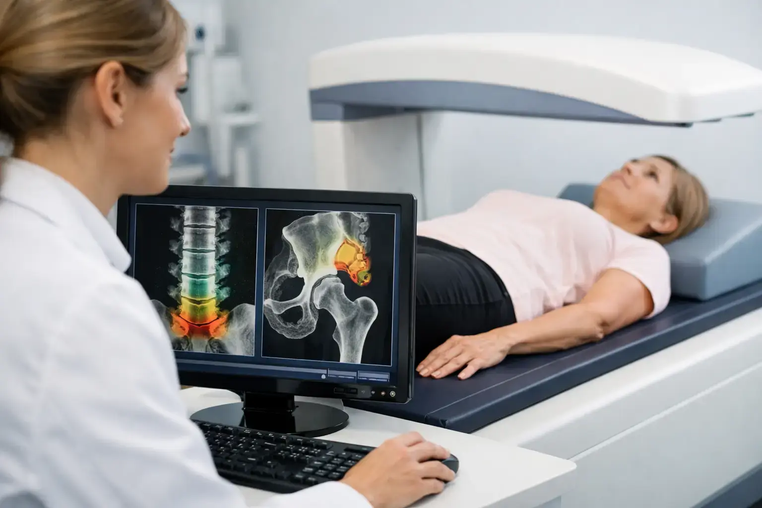
A cancer diagnosis brings a great deal of information, decisions, and follow-up. Bone health can become one more concern, especially when treatment affects hormones, ovarian function, nutrition, activity, or medication use. Cancer treatment bone loss screening is not about adding fear to an already demanding process. It is about making a largely silent change visible before a fracture is the first sign.
Bone loss is not inevitable for every person receiving cancer treatment, and the degree of risk depends on the cancer, treatment plan, age, sex, baseline bone health, and other medical factors. But the connection is established enough that bone health belongs in many cancer-care conversations.
Why cancer treatment can affect bone
Bone is living tissue. Throughout life, it is continually remodeled: specialized cells remove older bone while others lay down new mineralized bone. When these processes stay reasonably balanced, bone mass is maintained. When breakdown outpaces formation, bone mineral density can decline.
Several cancer treatments can shift that balance. Hormone-lowering therapies are a common example. Estrogen helps restrain bone resorption, so treatments that sharply reduce estrogen - including aromatase inhibitors used for some breast cancers and therapies that cause early menopause or ovarian suppression - can accelerate bone loss. Androgen-deprivation therapy for prostate cancer can have a similar effect through lowered testosterone and estrogen signaling.
Chemotherapy can also affect the bones indirectly when it causes premature menopause or lowers ovarian function. Long-term or repeated glucocorticoid use, sometimes part of treatment or supportive care, is another recognized contributor because these medications can alter bone remodeling. Stem cell transplantation, nutritional disruption, lower body weight, reduced mobility, and changes in vitamin D status may add to the picture.
That does not mean every treatment has the same skeletal effect. A person receiving a short course of treatment with stable hormone function has a different risk profile from someone beginning years of hormone suppression. The practical point is simpler: your bones may be responding to treatment even when they do not hurt.
What bone loss screening is trying to find
A fracture is a late event. Screening aims to identify risk earlier, when there is still time for your oncology team, primary care clinician, or bone-health specialist to interpret the pattern and discuss the appropriate next steps.
The standard starting point is usually bone mineral density, commonly measured at the lumbar spine and hip. Density is meaningful. It tells you how much mineralized bone is present, and it remains central to diagnosing osteoporosis and tracking change over time.
For cancer-treatment bone loss screening, a baseline measurement can be especially useful because it gives later results context. Without a baseline, a scan can describe where bone density stands now, but it cannot show how quickly it changed during treatment. The timing and frequency of follow-up testing should be determined by the clinical context, including the therapy being used and the initial result.
A bone-health conversation may also include fracture history, family history, menopause status, body weight, smoking, alcohol use, medical conditions, laboratory findings, and medications. Screening is most useful when a number is interpreted alongside the person who produced it.
Density matters, but it does not tell the whole story
Here is the limitation worth understanding: density measures quantity, not every signal associated with fragility.
Imagine two people whose hip density results fall in the same range. One may have no prior fractures and few additional risk factors. The other may be undergoing hormone-lowering cancer therapy, have lost bone quickly, and show ultrasound signal characteristics associated with greater skeletal fragility. Their density number may look similar, while their overall fracture-risk picture is not.
This is not an argument against density. It is an argument against asking density to answer more than it can. A density result cannot, on its own, fully represent the biological effects of cancer treatment, the pace of change, or all the factors that influence whether a fracture occurs.
Cancer care already works this way in other areas. A single lab value rarely tells the entire clinical story. Trends, treatment exposure, symptoms, and related measurements all matter. Bone health deserves the same level of attention.
What REMS adds to cancer treatment bone loss screening
REMS, or Radiofrequency Echographic Multi Spectrometry, uses radiation-free ultrasound to measure bone mineral density at the lumbar spine and proximal femur. It also analyzes raw ultrasound signals reflected from the bone to derive an independent Fragility Score.
That distinction matters. The Fragility Score is not another way of restating density. It reflects signal features associated with skeletal fragility that density alone does not capture. REMS then weighs density and the Fragility Score together to estimate five-year fracture risk.
A REMS report therefore addresses three related questions: how much mineralized bone is present, whether the reflected signal shows signs associated with greater fragility, and what those findings may mean together for estimated fracture risk over the next five years.
For someone with a cancer-treatment history, that broader view can be useful. A reassuring density result may still need context if treatment exposure or other risk factors suggest the skeleton deserves closer attention. Conversely, a low-density result becomes more informative when it is considered alongside a separate measure of fragility and a fracture-risk estimate.
REMS is FDA-cleared and does not use ionizing radiation. Its reported intra-operator precision error is below 0.5%, compared with typical reported DXA precision ranges of 0.9% to 1.9% at the spine. Precision is not a technical footnote. When bone can change relatively quickly, lower measurement error can help distinguish a likely real change from ordinary variation in the test.
That does not make one scan a prediction of your individual future. Fracture risk is influenced by falls, medications, age, prior fractures, treatment duration, and many other variables. It does mean the conversation can extend beyond a single density category.
When to raise the question with your care team
The right time to discuss screening is often before treatment begins, but it is also reasonable to raise the subject during treatment or years afterward. This is particularly relevant if your treatment lowers sex hormones, caused early menopause, includes glucocorticoids, or was accompanied by substantial weight loss or reduced mobility.
If you already have osteopenia or osteoporosis, the question changes slightly. Rather than asking only whether bone density is low, you may want to understand the trajectory: Has it changed? Is there other information about fragility? How does your current treatment history affect the interpretation of the result?
A previous low-trauma fracture deserves specific attention. So does new back pain, height loss, or a noticeable change in posture, since vertebral fractures can occur without a dramatic fall. These symptoms have many possible causes, particularly during or after cancer care, which is exactly why they warrant clinical evaluation rather than assumption.
Bring better information into a complicated conversation
Cancer treatment asks a lot of your body. Bone health should not be treated as an afterthought simply because bone loss is quiet. You do not need to wait for pain or a fracture to ask what treatment may mean for your skeleton.
A clear baseline, follow-up data when clinically appropriate, and an understanding of both density and fragility can give you and your care team a more complete place to begin. The goal is not to predict every outcome. It is to replace uncertainty with information you can carry into the next conversation.