
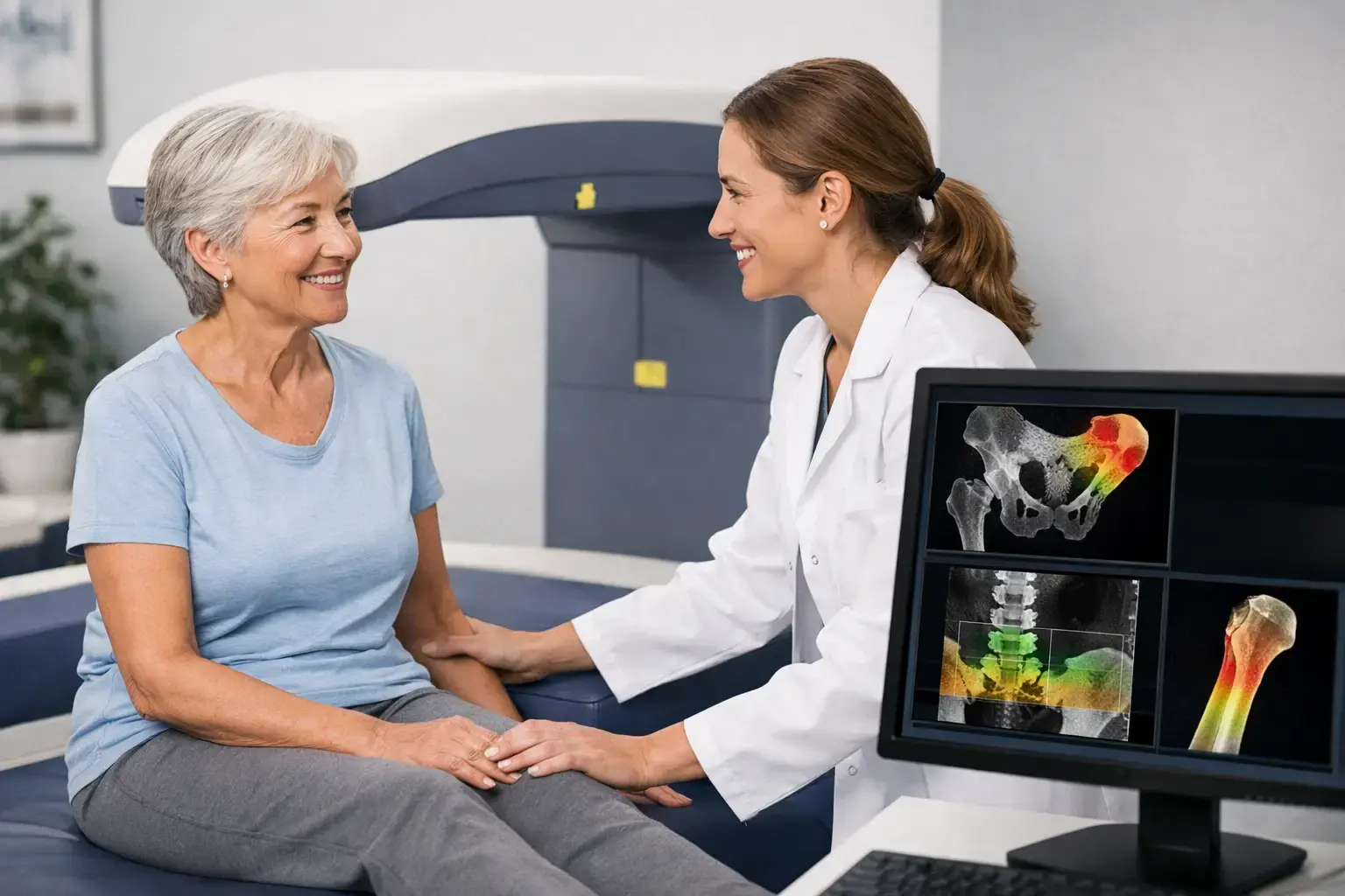
If you are entering menopause, managing osteopenia, taking a medication that affects bone, or simply trying to get ahead of preventable risk, “how often should bone density be tested?” is not a question with one calendar-based answer. The right interval depends on what is changing in your body, what the first scan showed, and whether the test can reliably detect a meaningful change.
A bone scan is not like an annual cholesterol panel. Bone usually changes gradually, and repeating a measurement too soon can create more noise than knowledge. But waiting years by default can also miss periods when bone is changing faster - or leave important questions unanswered when density alone appears reassuring.
The short answer: baseline first, interval second
For many adults, the most useful first step is a baseline assessment rather than choosing a repeat-testing schedule in advance. A baseline establishes how much mineralized bone is present at the lumbar spine and hip. It also gives future measurements something real to be compared against.
Routine population screening recommendations are generally age- and risk-based. In the United States, women age 65 and older are commonly recommended for osteoporosis screening. Younger postmenopausal women may be assessed earlier when risk factors are present. Guidance for men is less uniform, though many clinical organizations support assessment at older ages or earlier when risk is elevated.
That is a starting point for public-health screening, not a complete answer for an individual. Menopause, a prior low-trauma fracture, long-term glucocorticoid use, certain cancer treatments, rapid weight loss, and conditions affecting hormone status, nutrient absorption, or mobility can change the timing of assessment. So can a known diagnosis of osteopenia or osteoporosis.
For someone without a known risk factor and with a reassuring initial result, years may pass before another density measurement is likely to show a meaningful difference. For someone in a period of accelerated change or monitoring a clinical decision, a shorter interval may be reasonable. The point is not to scan on a fixed schedule. It is to measure when the result could add useful information.
Why the testing interval is not just about time
Bone is living tissue. It is continuously remodeled: older bone is resorbed and new bone is formed. Mechanical loading signals bone to adapt, while estrogen loss, some medications, illness, aging, and reduced loading can shift that balance toward loss.
The pace of that change varies. Bone loss can accelerate around the menopausal transition, for example, while a person may not meet routine age-based screening criteria for years. In other situations, changes may be slower enough that testing every year will not reveal a true biological shift.
There is another practical limit: every measurement tool has precision error. If a follow-up result changes by less than the amount the instrument can measure consistently, it may not reflect a real change in bone. It may be normal measurement variation.
This is why a repeat scan should be interpreted in terms of the least significant change - the amount of difference needed before clinicians can be confident that the result is more than noise. A shorter interval only becomes valuable when the method is precise enough and the clinical context suggests a meaningful change could occur.
When a one- to two-year follow-up may make sense
A one- to two-year interval is often discussed after an abnormal density result or during a period when bone status may be changing. It is not a universal rule, but it can provide a useful window for following a trend.
This timing may be considered when you have osteopenia or osteoporosis, have recently gone through menopause, have had a low-trauma fracture, or are navigating a treatment or health change known to affect bone. It can also be relevant after substantial weight loss or with long-term use of medications such as glucocorticoids, which can alter bone remodeling.
The reason for repeating the test matters as much as the interval. Are you establishing whether bone density is stable? Looking for evidence of ongoing loss? Bringing objective information to a conversation about medication? Each question calls for interpretation in context, rather than treating the next T-score as a pass-or-fail grade.
For people with stable results and no major change in risk, a longer interval is often appropriate. Testing more frequently does not automatically create better information. Good monitoring means measuring often enough to see a genuine signal, not often enough to generate a spreadsheet.
Density is necessary, but it does not tell the whole fragility story
Bone mineral density remains a central part of skeletal assessment. It measures how much mineralized bone is present, and low density is associated with fracture risk. But density is not the same thing as fragility.
Consider two people with similar density at the hip. One may have no other signals suggesting elevated fragility. The other may have a different pattern in the bone-related ultrasound signal that is associated with greater fragility. A density-only result may place them in the same category, even though their fracture-risk picture is not identical.
Type 2 diabetes makes this gap especially clear. People with type 2 diabetes can have bone density that appears normal or even higher than expected, yet research has consistently found higher fracture risk than density alone would predict. The biology is complex and can include changes related to chronic hyperglycemia and bone material properties that a density measurement does not directly capture. A normal density result is useful information, but it is not always the end of the conversation.
That distinction matters when deciding how often to test. If the only question is whether mineralized bone has changed, long intervals may be reasonable when change is slow. If the question is broader - how much bone is present, whether there are signals associated with fragility, and what the combined picture suggests about near-term fracture risk - the assessment itself needs to be broader.
What REMS adds to a follow-up plan
REMS, or Radiofrequency Echographic Multi Spectrometry, uses radiation-free ultrasound to measure bone mineral density at the lumbar spine and proximal femur. It also analyzes raw ultrasound signals reflected from bone to derive an independent Fragility Score.
That score does not replace density. It adds information beyond density, reflecting signal features associated with skeletal fragility. REMS then weighs density and Fragility Score together to estimate five-year fracture risk.
This gives a follow-up conversation more than one number. You can follow density over time, see whether the Fragility Score tells a similar or different story, and consider the estimated fracture risk in the context of your health history. For someone who has been told, “Your density is not that low,” but still has meaningful risk factors, that added layer may be particularly relevant.
Precision also affects timing. MobileREMS uses FDA-cleared REMS technology with an intra-operator precision error below 0.5%, compared with approximately 0.9% to 1.9% commonly reported for DXA at the spine. Lower measurement error means smaller changes may be distinguishable from machine variation. In settings where bone can change more quickly, that can make remeasurement over months, rather than years, scientifically meaningful.
That does not mean every person needs frequent scans. A precise measurement is most valuable when it answers a real question: Is there a measurable trend? Does a period of change appear to be affecting bone? Do density and fragility indicators point in the same direction?
Questions that make the interval clearer
Rather than asking only, “When is my next scan due?” it can help to bring a more specific set of questions to your clinician. What risk factors are present now? Has anything changed since the last assessment that could affect bone remodeling? Is the goal screening, monitoring, or informing a treatment decision? And does the testing method provide enough precision and enough information beyond density for that goal?
Your age matters, but it is only one part of the answer. A 52-year-old in menopause with rapid bone change may need a different monitoring conversation than a healthy 68-year-old with stable measurements. A person with type 2 diabetes and apparently normal density may need a fuller discussion of fracture risk than the density category suggests. Someone with established osteoporosis deserves careful trend monitoring without being reduced to a single T-score.
The best time to learn about your bones is before a fracture forces the question. A well-timed assessment can make an invisible process understandable, give you a baseline to follow, and help you have a more informed conversation about what your bones are showing now.