
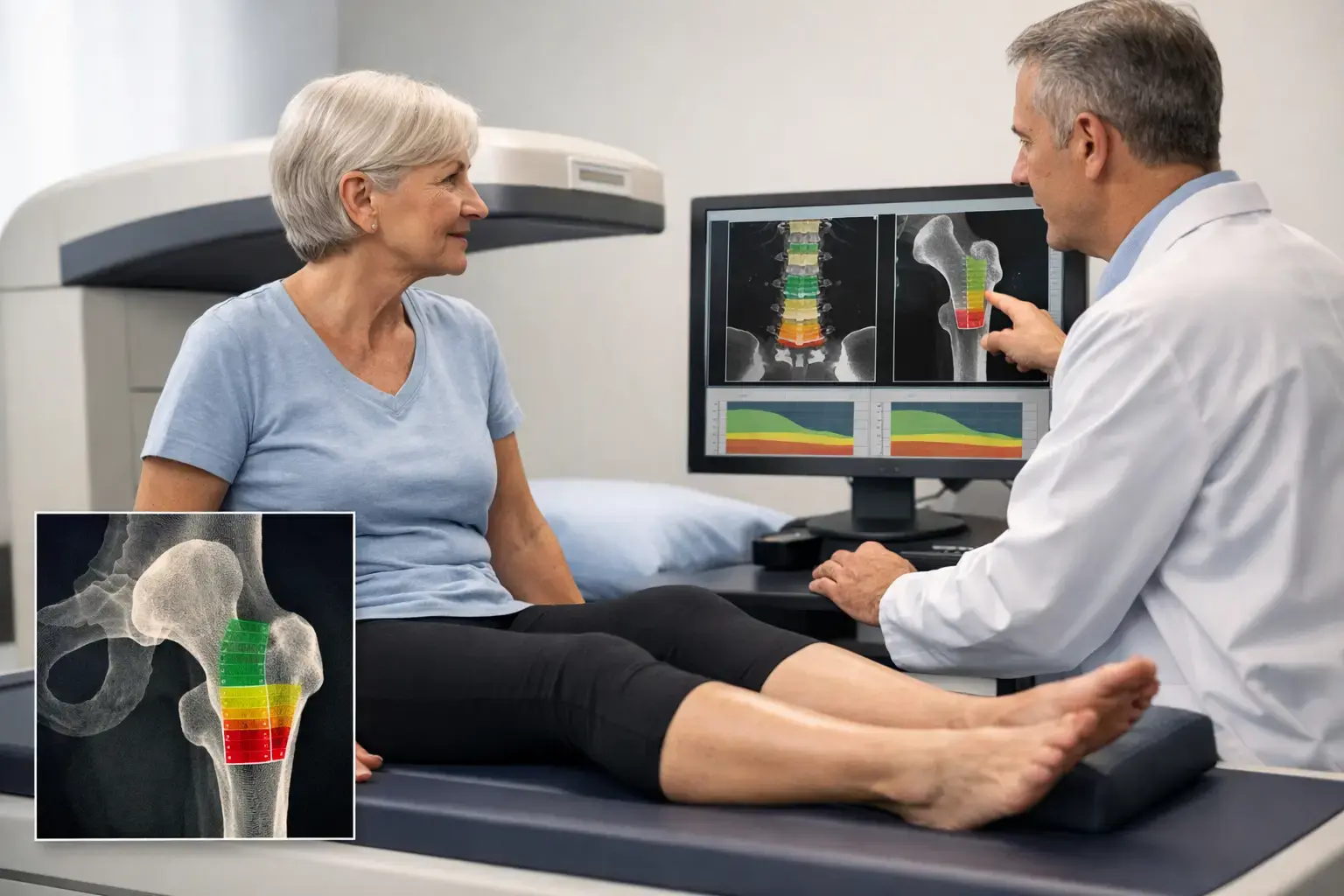
Prednisone can be essential treatment. It can also begin changing bone biology before you feel anything different, which is why a long term prednisone bone density test is not simply a routine checkbox. The useful question is broader: how much mineralized bone is present, are there signs of greater skeletal fragility, and what does that combination mean for fracture risk?
Glucocorticoid-related bone loss is often silent. A fracture, especially a vertebral compression fracture, may be the first visible consequence. The goal of testing is not to make a person taking prednisone afraid of a medication they may need. It is to make an otherwise invisible effect measurable early enough to inform a thoughtful conversation with their clinician.
Why prednisone affects bones
Prednisone is a glucocorticoid. These medications help control inflammation and immune activity, but their effects extend to the cells that continuously renew bone. Bone is living tissue, not a fixed frame. Throughout adulthood, old bone is removed and new bone is formed in a coordinated process called remodeling.
Glucocorticoids can disrupt that balance in several ways. They reduce the activity and survival of osteoblasts, the cells responsible for building bone, while early in treatment they can favor bone resorption. They can also affect calcium handling and alter sex-hormone signaling, both relevant to skeletal maintenance. The result can be less new bone formation at the same time the body remains vulnerable to loss.
That mechanism helps explain why glucocorticoid-associated fracture risk can rise relatively quickly. Bone mineral density may decline, particularly in the spine and hip, but density is not the entire story. The spine contains a high proportion of trabecular bone, the more metabolically active internal bone structure that can be especially affected by glucocorticoid exposure.
Dose, duration, underlying condition, age, menopause status, body weight, prior fractures, smoking, alcohol use, and other medications all influence the picture. A short course is not the same as months or years of treatment. Nor does every person taking long-term prednisone have the same level of risk.
When a long-term prednisone bone density test enters the conversation
Clinical guidelines commonly treat ongoing glucocorticoid use as a reason for early fracture-risk assessment. The American College of Rheumatology guidance addresses adults taking the equivalent of 2.5 mg or more of prednisone daily for more than three months, with the timing and scope of assessment shaped by the person's clinical circumstances.
In practice, a clinician may consider testing near the start of prolonged treatment rather than waiting for a conventional age-based screening milestone. That distinction matters. Many people first encounter bone-density testing because of age or menopause. Prednisone changes the reason for looking: the question becomes whether a medication-related risk is already affecting bone health.
A baseline is useful because it gives future measurements context. Without one, a later result can show where bone density stands but cannot reveal the direction or pace of change during treatment. If prednisone continues, repeat testing may be considered over time. The appropriate interval depends on the dose, duration, baseline findings, treatment changes, and the clinician's estimate of fracture risk.
Testing should also be discussed sooner when there has been a low-trauma fracture, unexplained loss of height, new persistent mid-back pain, early menopause, a history of eating disorders, conditions that affect nutrient absorption, or other osteoporosis risk factors. These situations do not establish a diagnosis by themselves. They add information that can change how a bone result is interpreted.
What standard density testing tells you
DXA, or dual-energy X-ray absorptiometry, remains the established standard for measuring bone mineral density and is central to most osteoporosis care pathways. It typically measures the lumbar spine and hip, producing a T-score that compares your density with the average bone density of a healthy young adult reference population.
That number matters. Lower density is associated with greater fracture risk, and a DXA result can identify osteopenia or osteoporosis according to established diagnostic thresholds. For people on prednisone, it can also provide an objective starting point for discussions about monitoring and fracture prevention.
But density is a quantity measurement. It tells you how much mineralized bone is present in the measured area. It does not, by itself, fully characterize every biological feature associated with fragility.
Consider two people with the same hip density score. One may have additional signals associated with greater skeletal fragility, while the other may not. Their density result looks similar, yet their fracture-risk picture may differ. This is not a reason to dismiss density. It is a reason to understand its boundary.
What else may belong in a prednisone bone assessment
A complete assessment is usually more than one scan result. A clinician may consider fracture history, height change, medication exposure, fall risk, family history, relevant laboratory findings, and whether vertebral imaging is warranted. Vertebral fractures are particularly easy to miss because they do not always produce a dramatic injury event.
Fracture-risk calculators can organize some of this information, though they also have limitations. Glucocorticoid exposure is often included as a risk factor, but risk may vary meaningfully by dose and duration. A calculation is an estimate, not a forecast written in stone.
For someone taking prednisone, the most useful result is therefore not always the lowest number on a report. It is the result placed in context: current density, evidence of change over time, clinical risk factors, and any signs that the skeleton may be more fragile than density alone suggests.
What REMS adds beyond density
REMS, or Radiofrequency Echographic Multi Spectrometry, measures bone mineral density at the lumbar spine and proximal femur using radiation-free ultrasound. It then analyzes the raw ultrasound signals reflected from bone to derive an independent Fragility Score.
The Fragility Score is not another T-score, and it does not diagnose osteoporosis on its own. It reflects signal patterns associated with skeletal fragility that bone mineral density alone does not capture. REMS weighs density and this independent fragility information together to estimate five-year fracture risk.
For a person on long-term prednisone, that distinction can be meaningful. Prednisone's effect on bone is not limited to the amount of mineralized bone measured at a single moment. A result that includes density, a Fragility Score, and an estimated five-year fracture risk offers three separate pieces of information to bring into a clinical discussion.
REMS does not replace every role of DXA or every component of a medical evaluation. DXA remains embedded in diagnostic criteria, treatment pathways, and many insurance processes. Rather, REMS can add information where the question is larger than density alone: Is this bone showing ultrasound signal characteristics associated with greater fragility, and how does that affect estimated fracture risk?
Its radiation-free design and reported intra-operator precision error below 0.5% also matter for longitudinal monitoring. A lower measurement error makes smaller changes easier to distinguish from ordinary test variation. Whether repeat measurement in months is useful depends on the clinical situation and should be interpreted alongside medication exposure and the rest of the bone-health picture.
Questions worth bringing to your appointment
If prednisone is expected to continue, a focused conversation can clarify more than a generic request for a scan. Ask how your dose and expected duration affect fracture risk, whether you need a baseline density measurement, and whether vertebral fracture assessment or spine imaging is relevant. It is also reasonable to ask what result would change the monitoring plan or prompt further evaluation.
If you already have a DXA result, ask what it does and does not answer in your case. A normal or mildly low density result can be reassuring, but reassurance is strongest when it matches the rest of your risk profile. For someone with ongoing glucocorticoid exposure, the absence of a severely low T-score does not automatically mean there is no bone-health question left to ask.
Prednisone may be necessary, and stopping or changing it is a medical decision that belongs with the clinician managing the condition it treats. Bone testing serves a different purpose: it gives you a clearer view of what the medication, your biology, and time may be doing to your skeleton.
Bones rarely announce change as it happens. A well-timed assessment turns that silence into usable information - before a fracture has to do the explaining.