
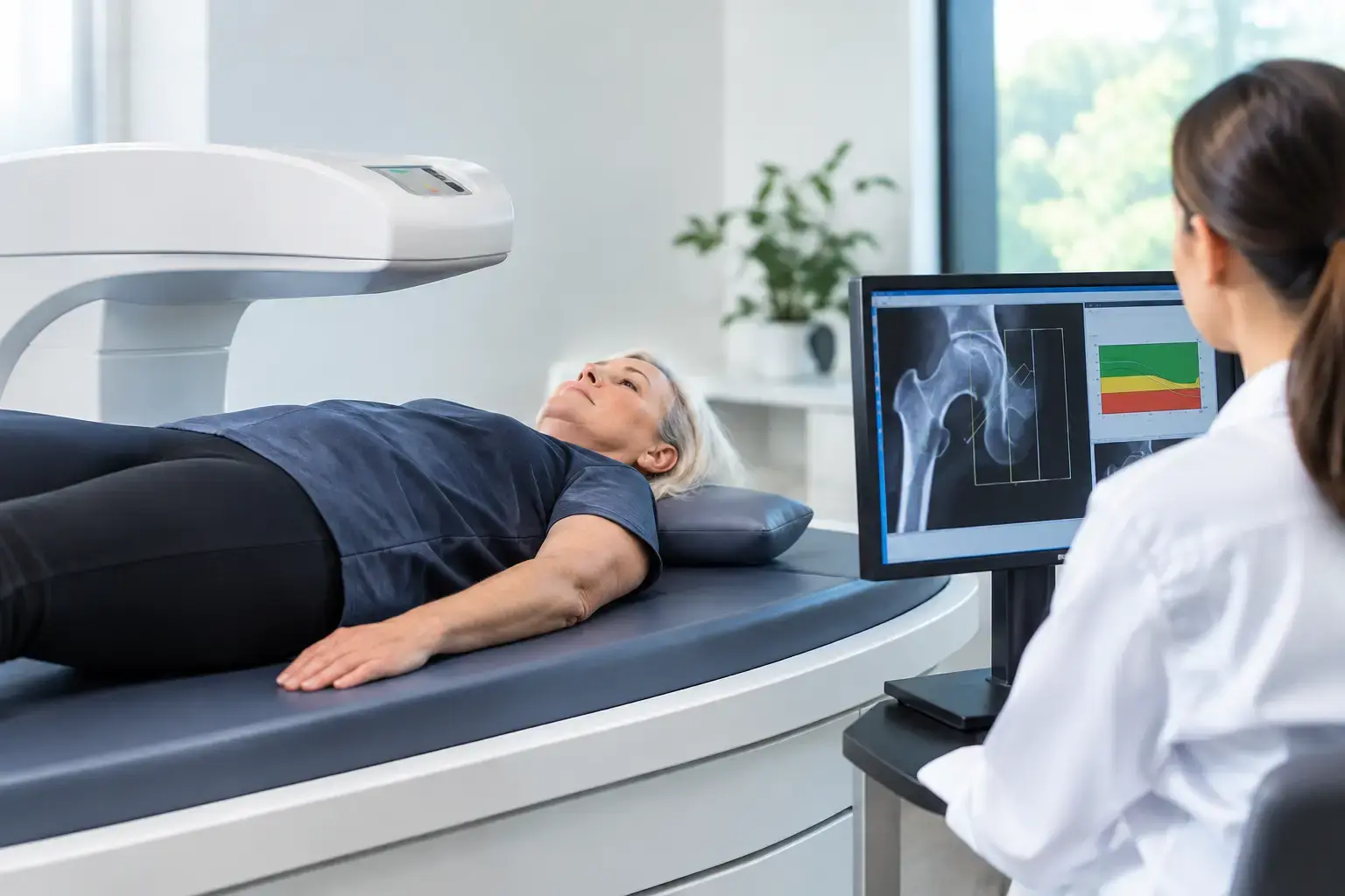
Your bones can be changing for years before anything hurts, breaks, or appears on a routine screening schedule. That is why learning how to build a bone baseline is less about predicting a problem and more about replacing guesswork with a starting point you can understand.
Many people have detailed data on cholesterol, glucose, body composition, and cardiovascular fitness. Yet they reach perimenopause, begin a medication that affects bone, lose weight quickly, or receive an osteopenia diagnosis without knowing two basic things: how much mineralized bone they have and whether that bone is showing signs associated with greater fragility.
A useful bone baseline answers both questions. It also gives future measurements context. A number taken five years from now means much more when you know where you started.
What a bone baseline is, and what it is not
A bone baseline is an objective snapshot of skeletal health at a particular point in time. It is not a verdict on your future, and it is not a promise that your bones will remain unchanged. Bone is living tissue. It responds to hormonal change, aging, mechanical loading, nutrition, illness, medications, and shifts in body weight.
The value of a baseline is comparison. If a later measurement changes, you and your clinician can ask whether the difference is likely to reflect a meaningful biological change or ordinary measurement variation. Without an earlier reference point, it is harder to tell whether a current result represents a long-standing pattern, recent loss, or stability.
The best baseline is broader than one density score. Bone mineral density, usually expressed through a T-score or Z-score, measures how much mineralized bone is present in the areas assessed. It remains a central part of bone evaluation. But density alone does not capture every signal related to skeletal fragility.
Think of two people whose density results are similar. One may have ultrasound signal characteristics associated with greater fragility, while the other does not. A density-only result may place them in the same category even though their overall fracture-risk picture is not identical. That gap is precisely why a baseline should include information beyond density when it is available.
Why timing matters before standard screening
Peak bone mass is largely built by the late twenties, and genetics influences much of the ceiling. From midlife onward, the question often shifts from how much bone you built to how well you are maintaining it and whether signs of fragility are appearing.
For women, the menopause transition is a particularly consequential period because declining estrogen can accelerate bone loss. Routine population screening, however, often begins later than the biological change. A baseline during perimenopause or early menopause can make a silent transition visible rather than leaving you to infer it years afterward.
Other circumstances can make earlier information especially useful. Long-term glucocorticoid use can affect bone remodeling. Cancer treatment histories may alter skeletal risk, although the specifics depend on the treatment and individual clinical context. Rapid weight loss can coincide with changes in bone, and people using GLP-1 medications may reasonably want objective bone information alongside metabolic and body-composition data.
Type 2 diabetes deserves particular attention because it illustrates the limits of density alone. Research has repeatedly found that people with type 2 diabetes can have normal or even higher bone mineral density while experiencing elevated fracture risk. The proposed biology is complex and may involve changes in bone material properties and accumulated effects of glycation, among other factors. The practical point is clearer: a reassuring density result does not always settle the question of fragility.
How to build a bone baseline that can be followed
A meaningful baseline begins with the measurement itself, but it should not end there. The report needs to provide information you can revisit with your physician as your health, medications, and life stage change.
Start with bone mineral density
Bone mineral density at the lumbar spine and proximal femur provides the established quantitative foundation. These are clinically relevant sites, and their results can be expressed in familiar terms such as T-scores and Z-scores. Density tells you how much mineralized bone is present. That matters, particularly when density is low.
Still, a low or normal density result is not the whole story. Treating the score as a complete portrait can create false reassurance in some people and unnecessary uncertainty in others. A baseline should make room for the distinction between bone quantity and signs associated with fragility.
Add independent information about fragility
REMS, or radiofrequency echographic multi-spectrometry, uses radiation-free ultrasound to assess bone mineral density at the lumbar spine and proximal femur. It also analyzes raw ultrasound signals reflected from bone to derive a Fragility Score.
The Fragility Score is independent of density. It reflects signal features associated with skeletal fragility that density alone does not capture. It does not directly measure flexibility, microarchitecture, failure load, or every biological contributor to fracture. Those distinctions matter. Its role is to add another clinically meaningful piece of information to the density result, not to claim that one measurement sees everything.
With REMS, density and Fragility Score can be weighed together to estimate five-year fracture risk. This gives a baseline three numbers that answer different questions: how much mineralized bone is present, whether the signal shows signs associated with greater fragility, and what the combination may mean for near-term fracture risk.
Make the result usable over time
A baseline only helps if later comparisons are trustworthy. This is where measurement precision becomes more than a technical detail.
Every measurement tool has some degree of variability. If that variability is large, a small difference between two reports may be noise rather than a real change in your bones. REMS has an intra-operator precision error below 0.5%, compared with the 0.9% to 1.9% typically reported for DXA at the spine. That tighter precision can support meaningful remeasurement over months rather than requiring years to see whether a reported change is likely to be real.
No technology eliminates the need for clinical interpretation. Changes in health status, medications, fracture history, and laboratory findings can all matter. But a precise measurement gives that conversation a stronger factual foundation.
Questions that make a baseline more useful
The most useful bone report is one you can explain in plain language. When reviewing any baseline with a clinician, ask what the density result shows, whether there is information about fragility beyond density, and how the available findings affect estimated fracture risk.
It is also reasonable to ask what future comparison would be meaningful and why. The answer depends on your life stage, medical history, reason for testing, and the precision of the method used. Someone monitoring a period of rapid biological change may have a different follow-up discussion than someone establishing a prevention-focused reference point.
Bring relevant context to the conversation. A prior low-trauma fracture, family history, diabetes, use of glucocorticoids, menstrual or menopausal history, cancer treatment history, and major weight change can all influence how a result is understood. A scan is valuable data, but it is most informative when connected to the rest of your health picture.
A baseline is information before consequences
A fracture can make bone health suddenly urgent. A baseline makes it possible to ask better questions before urgency chooses the timing.
For someone with osteopenia or osteoporosis, that may mean seeing whether density and Fragility Score tell a similar story or a more complicated one. For a person moving through menopause, it may mean documenting a starting point during a period when bone can change quickly. For the health-focused person who tracks nearly everything except skeletal health, it may mean finally including the framework that carries them through every run, lift, walk, and ordinary day.
MobileREMS provides this broader view through radiation-free REMS assessment: bone mineral density, an independent Fragility Score, and an estimated five-year fracture risk. The goal is not to make your bones a source of fear. It is to make an invisible process understandable enough to follow, discuss, and act on with clear information when decisions arise.
Your bones do not need to wait for a consequence to become worth measuring.