
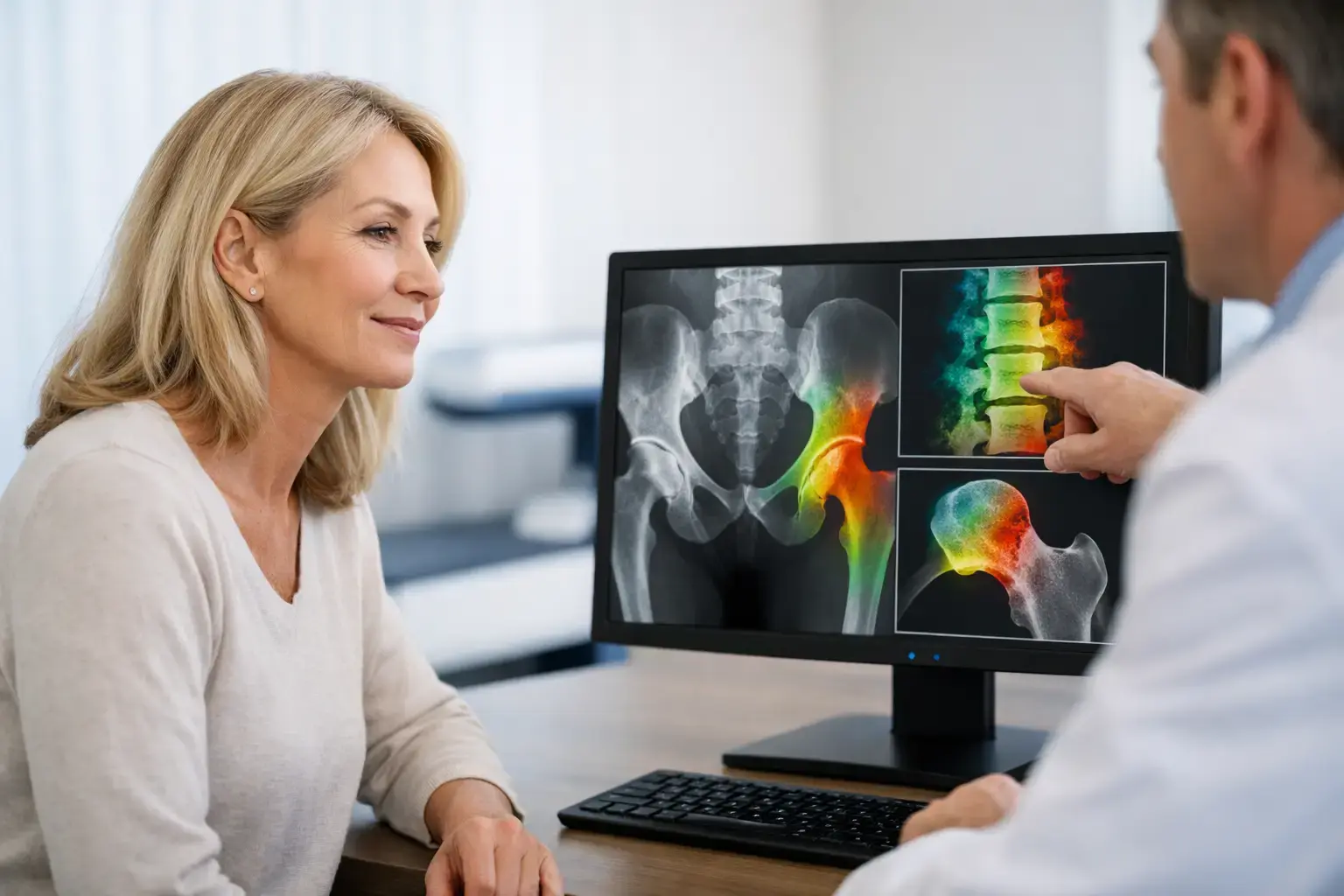
Menopause can change your bones long before anything hurts. If you are asking how to protect bones in menopause, the useful question is not only how to slow bone loss. It is also how to understand what is changing, establish a meaningful baseline, and avoid letting a fracture become the first sign that your skeleton needed attention.
Bone loss around menopause is common, biologically understandable, and not a personal failure. The transition marks a shift in the hormone signals that have helped regulate bone remodeling for decades. That shift can be rapid, particularly in the years surrounding the final menstrual period. The good news is that bone health is measurable, and meaningful information is available before consequences force the conversation.
Why menopause can accelerate bone loss
Your skeleton is living tissue. Throughout adulthood, specialized cells continuously remove small amounts of older bone and replace it with new bone. This remodeling process repairs microscopic damage and helps keep mineral balance in the body.
Estrogen is one of the hormones that helps keep this cycle in balance. As estrogen levels become more variable in perimenopause and then decline after menopause, bone resorption can outpace bone formation. In plain terms, more bone may be removed than rebuilt. The result can be a measurable decline in bone mineral density, especially at the lumbar spine and hip.
The pace is not identical for everyone. Genetics, the amount of bone built earlier in life, body weight changes, nutrition, activity, smoking, alcohol exposure, certain medications, medical conditions, and the timing of menopause all matter. An early menopause or surgical removal of the ovaries can mean less lifetime estrogen exposure and may change the risk conversation.
This is why a person who feels healthy, exercises regularly, and has no fracture history can still be losing bone during the menopausal transition. Bone loss is usually silent. A fracture is not.
How to protect bones in menopause: the foundations
Protecting skeletal health is not one habit or supplement. It is a set of inputs that influence bone remodeling, muscle function, balance, and fall risk over time.
Mechanical loading is one of those inputs. Bone responds to forces placed on it by adapting its structure and mineral content. Weight-bearing movement and resistance-based activity create loading signals that differ from non-weight-bearing exercise. Muscle strength also matters because muscles pull on bone, and because strength and balance can influence the likelihood of a fall. The most appropriate form of activity depends on prior injuries, fracture history, fitness, joint health, and whether osteoporosis is already present.
Nutrition provides another part of the equation. Calcium is a mineral stored in bone, while vitamin D supports calcium absorption and participates in bone metabolism. Adequate protein supports muscle and the bone matrix itself. Whether food intake, laboratory testing, or supplementation is relevant is an individual clinical question, particularly for people with kidney disease, gastrointestinal conditions, a history of stones, or medications that affect mineral metabolism.
Avoiding tobacco exposure and limiting heavy alcohol use also supports bone health. These are not magic levers, and none can guarantee that bone density or fracture risk will remain unchanged. They are modifiable influences within a larger biological picture.
Menopausal hormone therapy may be part of the discussion for some people. It can reduce bone loss and fracture risk while being used, but its use is not solely a bone decision. The balance of potential benefits and risks depends on age, time since menopause, symptoms, cardiovascular and breast cancer history, clotting risk, and other individual factors. A clinician who knows your history can place bone considerations in that broader context.
A density score matters, but it is not the whole story
A DXA scan has long been the standard way to measure bone mineral density. The T-score from that scan compares your density with the average density of a healthy young adult reference population. It is valuable information and remains central to diagnosing osteopenia and osteoporosis.
But density answers one question: how much mineralized bone is present? It does not fully answer whether that bone is showing signs associated with greater fragility.
Consider two people with similar bone density at the hip. One may have ultrasound signal characteristics associated with lower skeletal fragility, while the other may have characteristics associated with greater fragility. If you look only at density, their stories may appear similar. Fracture risk is not determined by density alone, however. Age, prior fracture, falls, medications, health conditions, and features of bone fragility all contribute.
This distinction matters during menopause because the goal is not simply to obtain a label. It is to understand your current skeletal position and make better-informed conversations possible. A normal or mildly reduced density result can be reassuring, but it should be interpreted in context rather than treated as a complete clearance signal.
When earlier bone information makes sense
Routine population screening often begins later than the menopausal transition. Yet risk can begin changing years before then. Earlier assessment may be worth discussing when menopause occurs early, periods stop after cancer treatment, there is a parent with a hip fracture, or there has been a low-trauma fracture.
Other reasons include long-term glucocorticoid use, conditions that affect nutrient absorption, rheumatoid arthritis, substantial unintentional weight loss, or medications and treatments known to affect bone. Type 2 diabetes deserves particular attention: bone density may be normal or even higher than expected, while fracture risk can still be elevated. Changes in bone material properties and a greater likelihood of falls may help explain this false reassurance from density alone.
A baseline does not predict your entire future. It gives future measurements a reference point. That is especially useful when the biological transition is active and decisions about lifestyle, medications, or hormone therapy may be under consideration.
What REMS adds to the menopause bone-health conversation
Radiofrequency Echographic Multi Spectrometry, or REMS, is a radiation-free ultrasound method that measures bone mineral density at the lumbar spine and proximal femur. It also analyzes raw ultrasound signals reflected from bone to derive an independent Fragility Score.
That score is not another density number. It reflects signal features associated with skeletal fragility that density alone does not capture. REMS then weighs density and Fragility Score together to estimate five-year fracture risk. The result is three understandable pieces of information: density, a Fragility Score, and an estimated fracture-risk measure.
For someone in perimenopause or menopause, that broader view can clarify an incomplete picture. A person with lower density may learn that the fragility signal and estimated risk align with the density concern. Another person with a less concerning density result may see information that supports a more careful conversation with their physician. Neither outcome is a diagnosis by itself, and neither replaces clinical judgment. Both are more informative than assuming density tells the entire story.
REMS is FDA-cleared and does not use ionizing radiation. Its low measurement error also supports following bone information over shorter intervals when clinically appropriate. Precision matters because a small apparent change is only useful if it is likely to represent a real biological change rather than ordinary measurement variation.
Bring the right questions to your bone-health visit
If you are nearing menopause, in the transition, or years beyond it, ask what your individual risk factors mean for timing and type of assessment. Ask whether a prior fracture was considered low trauma, whether medications or health conditions affect your bones, and what information beyond density would help clarify your fracture risk.
If you already have a T-score, ask what it does and does not explain. A number can be useful without being complete. The most productive bone-health conversation connects your density with your age, health history, fracture history, and evidence of fragility.
Menopause is a meaningful time to look at the skeleton because change can be happening quietly. You do not need to wait for pain or a fracture to make your bones worth knowing.