
A bone-density result can look reassuring and still leave a meaningful question unanswered: what does that number say about your likelihood of fracture? This osteoporosis test options review looks beyond the familiar scan to explain what each assessment can measure, what it cannot, and why those distinctions matter before a fracture forces the conversation.
For decades, bone mineral density has been the central measurement in bone health. It remains useful. But density measures the amount of mineralized bone present, not every signal associated with skeletal fragility. Two people can have similar density results and different fracture risk profiles, particularly when other factors affect bone quality or fall risk.
The right test depends on the question being asked. Are you establishing a baseline during menopause? Following diagnosed osteopenia? Looking for more clarity after a normal density result in the setting of type 2 diabetes, glucocorticoid exposure, rapid weight loss, or cancer treatment? A good assessment starts by matching the tool to the information gap.
Osteoporosis Test Options Review: What Each Test Measures
DXA: the established density test
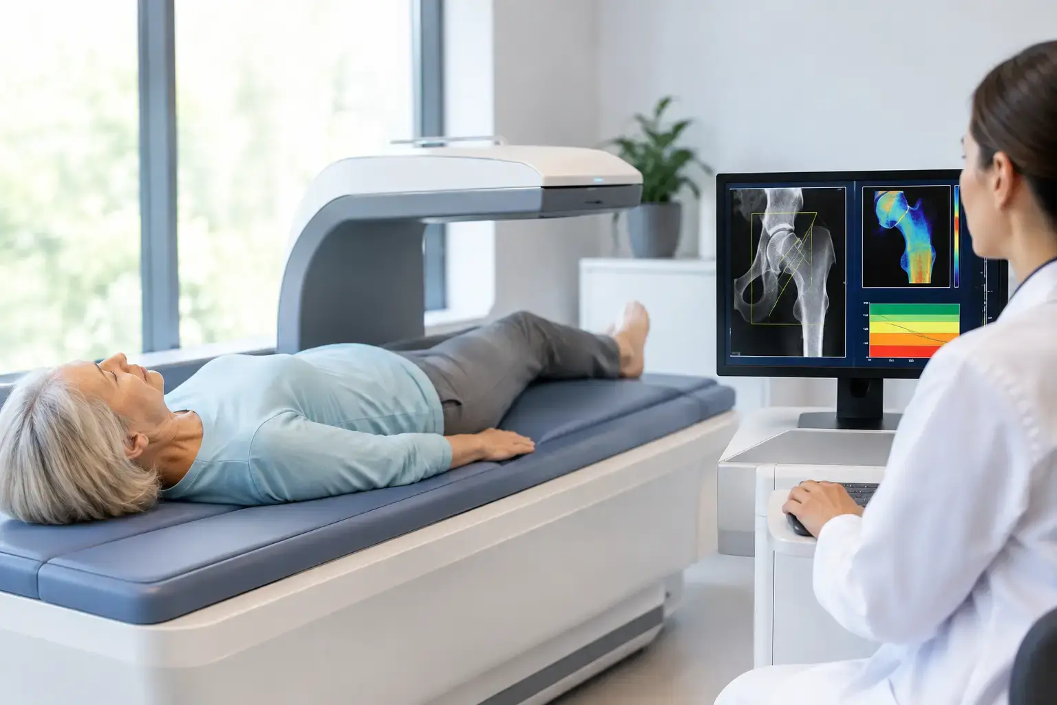
Dual-energy X-ray absorptiometry, usually called DXA, is the standard test used to diagnose osteoporosis. It measures bone mineral density, most commonly at the lumbar spine and hip. Those measurements are reported as T-scores, which compare your density with the average peak bone density of a young healthy reference population.
A T-score of -2.5 or lower meets the densitometric definition of osteoporosis. A score between -1.0 and -2.5 is generally classified as low bone mass, often called osteopenia. These categories are clinically useful, especially for establishing a common language between patients and clinicians.
DXA is widely available, relatively quick, and supported by decades of research. It is also the reference method used in many treatment guidelines and fracture-risk calculations. Its limitation is not that it measures density poorly. Its limitation is that density is one part of fracture risk.
DXA uses a small amount of ionizing radiation. For most people, the exposure is low, but it remains a consideration for repeated measurements and for people who prefer a radiation-free approach. Precision also varies by site, equipment, positioning, and facility. When comparing scans over time, consistency in the testing center and technique matters.
REMS: density plus an independent fragility signal
Radiofrequency Echographic Multi Spectrometry, or REMS, uses ultrasound rather than X-rays to measure bone mineral density at the lumbar spine and proximal femur. Those are the same clinically important skeletal sites commonly assessed with DXA.
What makes REMS distinct is what happens after the density measurement. The technology analyzes raw ultrasound signals reflected from the bone and derives a Fragility Score. This is an independent signal associated with skeletal fragility, not another name for bone density and not a direct measurement of bone microarchitecture, flexibility, or failure load.
That distinction matters. Density answers, “How much mineralized bone is present?” The REMS Fragility Score adds, “Is this reflected ultrasound signal showing patterns associated with greater fragility?” REMS then weighs density and the Fragility Score together to estimate five-year fracture risk.
Consider two people with similar hip density. Density alone places them in the same general category. If one has a higher Fragility Score, the fuller picture may be different. That does not mean a single score predicts an individual future with certainty. It means the assessment has identified information that density alone does not supply.
REMS is FDA-cleared and radiation-free. Its reported intra-operator precision error is below 0.5%, compared with the approximately 0.9% to 1.9% commonly reported for DXA at the spine. Precision is not an abstract technical advantage. It helps determine whether a change between measurements is likely to represent a real biological change rather than ordinary measurement variation.
That can be particularly relevant when bone status may be changing over months, not only years. MobileREMS uses this technology to provide density, a Fragility Score, and an estimated five-year fracture risk in one report that can be shared with your physician.
Quantitative CT: a three-dimensional density measurement
Quantitative computed tomography, or QCT, estimates volumetric bone mineral density in three dimensions. Unlike DXA, which reports an areal density measurement, QCT can distinguish trabecular bone from cortical bone in some applications. Trabecular bone is the more metabolically active, lattice-like bone found in areas such as the vertebrae.
QCT can be valuable in selected clinical situations, including when degenerative changes, spinal hardware, or body size make DXA interpretation more difficult. It may also be performed opportunistically from a CT scan obtained for another reason, depending on the scan and analysis available.
The trade-offs are radiation exposure, access, cost, and less standardization across settings than DXA. QCT is not usually the first-line population screening test, but it can answer a different question when three-dimensional information is clinically relevant.
Heel ultrasound: useful screening, limited comparison
Some devices use ultrasound at the heel, or calcaneus, to assess properties associated with bone status. Heel ultrasound is portable, radiation-free, and can be useful for community screening or identifying people who may benefit from further evaluation.
It does not measure the hip or lumbar spine, where osteoporosis is typically diagnosed by DXA or assessed by REMS. Results from heel ultrasound should not be treated as interchangeable with a central DXA T-score. A concerning result may be a reason to pursue a more complete central-site assessment; a reassuring result does not answer every question about spinal or hip fragility.
Vertebral fracture assessment: looking for silent fractures
A vertebral fracture assessment, often performed on a DXA system, uses low-dose imaging to look for compression fractures in the spine. This is a different task from measuring density.
Many vertebral fractures are not recognized when they happen. Back pain may be attributed to age, posture, or an old injury, and some fractures cause little obvious pain. Yet the presence of a vertebral fracture is clinically significant because a prior fragility fracture changes the conversation about future risk, regardless of whether density falls neatly into a diagnostic category.
A clinician may also use standard spine X-rays or other imaging when symptoms, height loss, posture changes, or medical history raise a specific concern.
When Density Can Miss Part of the Story
Type 2 diabetes is the clearest example of why density should not be confused with complete fracture-risk information. People with type 2 diabetes may have bone mineral density that is normal or even higher than expected, while fracture risk remains elevated. Research has repeatedly shown this mismatch.
The reasons are complex. Diabetes may affect collagen within bone through advanced glycation end products, alter bone turnover, and increase the likelihood of falls through factors such as neuropathy, vision changes, or some medications. A density result can therefore be real and still incomplete.
The same principle applies, though not identically, in other situations. Long-term glucocorticoid use can affect bone remodeling. Menopause can accelerate bone loss as estrogen declines. Rapid weight loss may change mechanical loading and nutritional status. Cancer therapies may affect bone through several pathways, depending on the treatment. No single test settles every one of these questions, but more relevant information supports a more informed discussion.
Tests That Support Bone Evaluation but Do Not Diagnose Osteoporosis
Blood and urine tests can help clinicians investigate contributors to low bone density or unexpected bone loss. Depending on the situation, this may include calcium, vitamin D status, kidney function, thyroid markers, parathyroid hormone, or markers of bone turnover.
These laboratory values answer different questions from an imaging-based bone assessment. Vitamin D participates in calcium absorption and bone metabolism, for example, but a vitamin D result does not tell you your hip density, Fragility Score, or fracture risk. Bone turnover markers can reflect remodeling activity, yet they do not independently diagnose osteoporosis.
Clinical fracture-risk calculators also add context. They combine age, sex, body size, prior fracture history, smoking, alcohol exposure, glucocorticoid use, certain medical conditions, and sometimes femoral-neck density. They are useful population-based estimates, but their output depends on the variables included. They do not replace an assessment designed to examine the bone directly.
Choosing the Information You Need
If you need a standard diagnostic density measurement that aligns with most guidelines, DXA remains foundational. If a clinician needs three-dimensional information or DXA is difficult to interpret, QCT may have a role. If the goal is quick screening, heel ultrasound may help identify who needs further evaluation.
If you want to understand density alongside an independent signal of fragility and a five-year fracture-risk estimate, REMS addresses a broader information gap at the spine and proximal femur without radiation. It is especially relevant when a T-score alone feels like an incomplete answer, not because density is unhelpful, but because your bones deserve more than one number.
The most useful bone test is the one that makes the invisible more understandable and gives your care team clearer information for the decisions in front of you. Before consequences become the first signal, it is reasonable to ask what your current measurement can see - and what it cannot.