
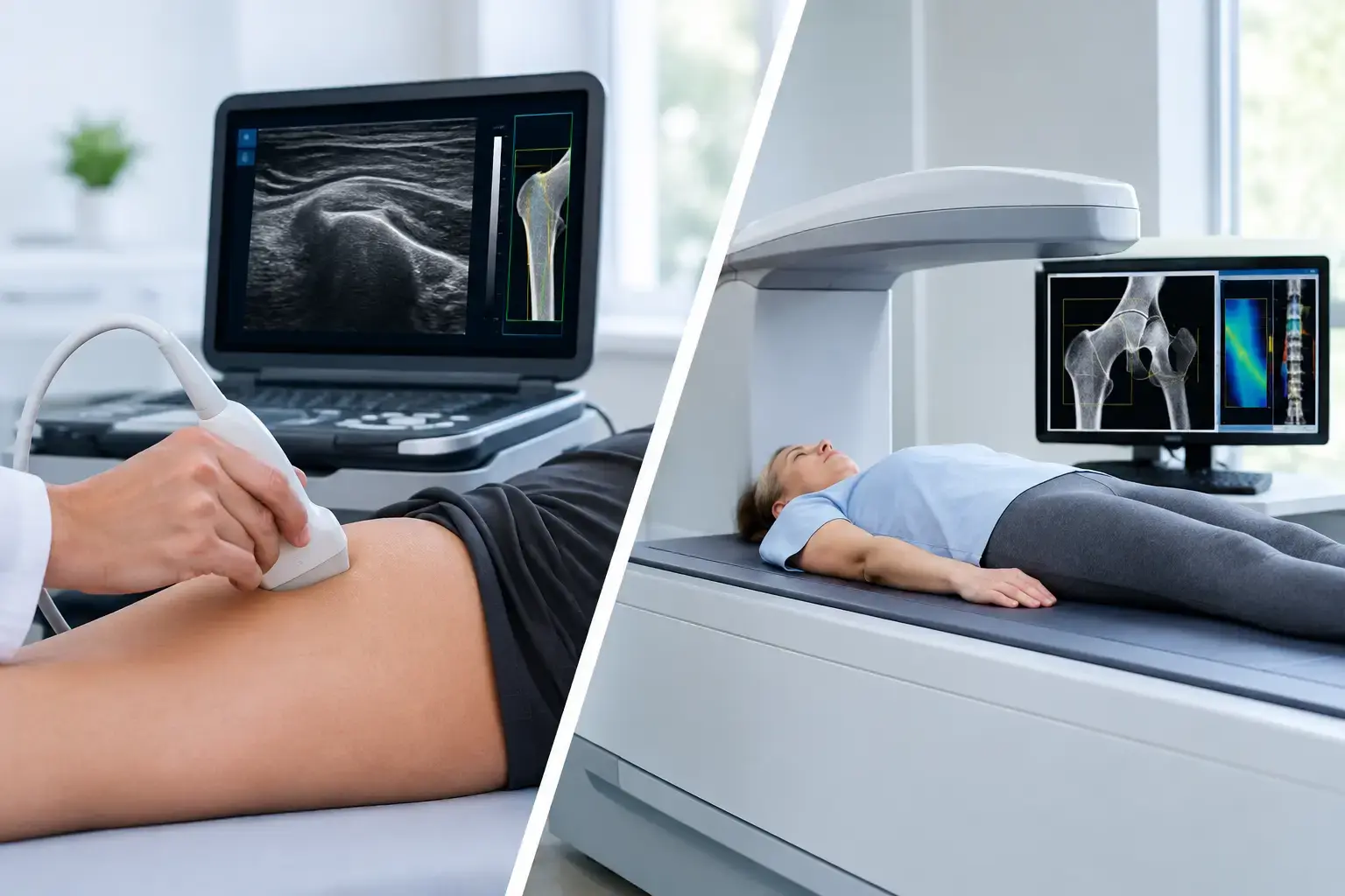
A bone-density result can look reassuring and still leave an important question unanswered: does your bone show signs associated with greater fragility? That is the real issue behind ultrasound versus DXA. The comparison is not simply about choosing an older test or a newer one. It is about deciding how much information you want before a fracture forces the conversation.
For decades, DXA has been the familiar answer to bone assessment. It remains an important clinical tool, particularly for diagnosing osteoporosis and tracking bone mineral density over time. But density is one part of skeletal health, not the whole of it. Two people can have similar density measurements and carry meaningfully different fracture risk.
Ultrasound Versus DXA: They Measure Different Things
DXA, short for dual-energy X-ray absorptiometry, uses two low-dose X-ray beams to estimate bone mineral density, or BMD. It is commonly performed at the lumbar spine and hip, the sites most relevant to osteoporosis assessment. The result is often reported as a T-score, which compares your density with the average density of a healthy young adult reference population.
That number matters. Lower bone mineral density is associated with higher fracture risk, and DXA has a long clinical history behind it. A DXA result can identify osteoporosis by established diagnostic thresholds and can give your physician a familiar baseline for clinical decision-making.
But BMD is fundamentally a measure of how much mineralized bone is present. It does not directly tell you whether the reflected characteristics of that bone are more or less associated with fragility.
REMS, or Radiofrequency Echographic Multi Spectrometry, approaches the question differently. It uses radiation-free ultrasound to measure BMD at the lumbar spine and proximal femur. Then it analyzes the raw ultrasound signals reflected from bone to derive an independent Fragility Score.
The distinction is meaningful. A REMS scan provides density, as well as information from the reflected signal that is associated with skeletal fragility. It then weighs those two measures together to estimate five-year fracture risk.
A simple way to picture the difference is to imagine two people leaving their scans with the same BMD. Density tells us they have a similar amount of mineralized bone. REMS may show that one person has a higher Fragility Score than the other. Their density tells a similar story; the signal-derived fragility information does not. That difference can change how fully they understand their fracture-risk picture.
Why a Normal Density Result Is Not Always the End of the Story
Type 2 diabetes is one of the clearest examples of why density alone can be incomplete. People with type 2 diabetes may have normal or even higher BMD, yet research has consistently found an elevated fracture risk. The reasons are complex and may involve changes in bone material properties and the broader effects of diabetes on falls, healing, and skeletal biology. The central point is straightforward: a normal density result does not automatically mean normal fracture risk.
This is not limited to diabetes. Fragility risk can become more complicated in people with long-term glucocorticoid exposure, a history of cancer treatment, rapid weight loss, or other health changes that affect bone. During perimenopause and menopause, bone loss can accelerate while routine screening may still feel distant. Someone may have no fracture, no obvious symptom, and no reason to suspect that their skeletal picture is changing.
Bone loss is often silent. The fracture is what makes it visible. Meaningful assessment creates an opportunity to see more of the process before that point.
DXA can be part of that assessment. The limitation is not that DXA is unhelpful. The limitation is treating one density number as though it answers every question about fragility.
What REMS Adds to a Bone Assessment
Not every ultrasound bone test is the same. Some ultrasound devices assess peripheral sites such as the heel and may be used as screening tools. REMS is different: it measures BMD at clinically relevant central skeletal sites, the lumbar spine and proximal femur, while analyzing the radiofrequency signals generated during the scan.
That analysis produces three results designed to be followed over time:
- Bone mineral density at the spine and hip
- An independent Fragility Score derived from reflected ultrasound signals
- An estimated five-year fracture risk that combines density and fragility information
The Fragility Score is not a second version of the T-score. It is independent information. It does not diagnose flexibility, brittleness, failure load, or every aspect of bone structure. It identifies signal characteristics associated with fragility, adding context that density alone does not provide.
That boundary matters because clear information is more useful than exaggerated claims. A REMS scan does not replace a physician’s clinical judgment, medical history, medication review, or evaluation of fall risk. It does offer a broader data set for those conversations.
For someone who has been told they have osteopenia or osteoporosis, that can mean moving beyond the understandable question, “How low is my T-score?” The next question may be, “Does the information about fragility tell the same story as my density?” For someone without a diagnosis, the question may be earlier and simpler: “What is happening in my bones before there is a consequence?”
Precision Changes What Monitoring Can Mean
The usefulness of any repeat measurement depends on whether it can distinguish a real biological change from ordinary measurement variation. This is where precision becomes practical rather than technical.
REMS is FDA-cleared and radiation-free, with an intra-operator precision error below 0.5%. Reported DXA precision at the spine is commonly in the range of 0.9% to 1.9%, though results vary by equipment, site, positioning, and facility protocol. Neither number tells the whole clinical story by itself, but lower measurement error can make smaller changes easier to interpret.
Why does that matter? Bone does not always change on a convenient annual schedule. Perimenopausal transition, medication changes, rapid weight loss, recovery from illness, and other physiological shifts can create reasons to want a clearer view sooner. When a test has tighter repeatability, it becomes more feasible to re-measure over months and have greater confidence that a change on the report reflects your bones rather than scanner noise or positioning differences.
Radiation exposure is also part of the practical comparison. DXA uses a very low dose of ionizing radiation. For many people, that exposure is not a barrier and is appropriately weighed against the value of the information obtained. REMS uses ultrasound rather than ionizing radiation, so radiation does not limit repeat assessment.
When DXA Still Has an Important Role
A useful ultrasound versus DXA discussion is not a contest with one universal winner. DXA remains the conventional reference method for BMD-based osteoporosis diagnosis, and many healthcare systems, insurers, and treatment pathways are built around DXA reports and T-scores. If your clinician needs a DXA result for a specific diagnostic, coverage, or treatment decision, that practical reality matters.
DXA can also be valuable when prior DXA studies provide a long-standing trend line. Comparing measurements from the same method, on the same equipment when possible, is often useful for interpreting change over time.
REMS is especially relevant when you want BMD at the spine and hip without radiation, along with independent fragility information and a five-year fracture-risk estimate. It may also be useful when the question is not merely whether density crosses a diagnostic threshold, but whether density tells the whole story for your particular risk profile.
The strongest approach is often not choosing a side. It is understanding what each measurement can tell you, what it cannot tell you, and how the result fits with the rest of your health information.
A More Complete Starting Point for Bone Health
Most people know their cholesterol, blood pressure, or glucose long before they know anything specific about their bones. Yet peak bone mass is largely established by early adulthood, genetics influences the ceiling, and bone loss may begin quietly well before a person considers themselves at risk.
Mechanical loading stimulates bone adaptation. Nutrition, hormones, health conditions, medications, body weight, and aging all influence skeletal biology. No scan captures every one of those factors. A well-chosen scan can make the invisible part more understandable.
If you are making sense of osteopenia, entering menopause, living with type 2 diabetes, monitoring a major health transition, or simply building a more complete health baseline, the question is not whether density matters. It does. The better question is whether density alone gives you enough information to understand your bones before consequences do.