
A missed period is visible. The menopausal bone changes that can begin during the same transition are not. If you are in perimenopause, recently postmenopausal, or simply trying to understand what this stage means for your long-term health, the useful question is not only whether bone loss is happening. It is whether you have enough information to see it before a fracture forces the conversation.
Bone is living tissue, continuously renewed through remodeling. Menopause changes the hormonal conditions under which that remodeling occurs, often quickly. Bone mineral density remains a central part of the picture, but it does not tell the entire story of skeletal fragility or fracture risk.
Why menopausal bone changes can accelerate
Estrogen helps regulate the balance between two cell types involved in bone remodeling: osteoclasts, which break down old bone, and osteoblasts, which form new bone. In the years around menopause, estrogen levels become more variable and then decline. Osteoclast activity can rise, while bone formation does not always keep pace.
This is not a switch that flips on the day periods stop. Perimenopause can last years, and bone loss may begin while cycles are still present but irregular. The pace varies substantially from person to person. Genetics, peak bone mass built earlier in life, body weight changes, smoking, alcohol exposure, certain medications, nutrition, physical activity, and medical conditions all influence the trajectory.
The result is often a period of faster net bone loss around the final menstrual period and in the early postmenopausal years. Trabecular bone, the lattice-like bone found in areas such as the vertebrae, is metabolically active and may be particularly affected. The hip also matters: hip fractures can carry serious consequences for mobility and independence, which is why the proximal femur is a key site in bone assessment.
None of this means a person in menopause is destined for osteoporosis or fracture. It does mean that waiting for symptoms is not a useful strategy. Bone loss itself is typically silent.
The number most people know: bone density
A bone mineral density test estimates how much mineralized bone is present at a measured site. Results are commonly expressed as a T-score, which compares your density with the average peak bone density of a healthy young adult reference population. A T-score can identify low bone mass and osteoporosis within established diagnostic thresholds.
That information is valuable. It can establish a baseline, reveal change over time, and help frame conversations with a clinician. But density is one dimension of bone health, not a complete measure of how likely a bone is to fracture.
Consider two postmenopausal women with similar bone density at the hip. One may have a fracture history, diabetes, long-term glucocorticoid exposure, or ultrasound signal features associated with greater skeletal fragility. The other may not. A density number alone can make those two situations look more alike than they are.
This is why a reassuring density result is not always the end of the question. Type 2 diabetes is the clearest example. People with type 2 diabetes can have normal or even higher bone mineral density while their fracture risk remains elevated. Changes related to diabetes can affect bone material and remodeling in ways density does not fully capture. Menopause can add another source of skeletal change to that already more complicated risk picture.
Menopausal bone changes are more than a density story
Fractures occur when force meets a bone with insufficient resistance for that event. Bone mineral density contributes to resistance, but it does not fully explain it. Clinical history matters, including age, prior fractures, falls, family history, medications, and conditions that affect bone. Signals associated with fragility matter as well.
This distinction is especially relevant during menopause because the transition can be biologically active long before a routine screening schedule begins. In the United States, standard population screening has traditionally focused on women age 65 and older, with earlier assessment considered for younger postmenopausal women who have risk factors. That approach is useful for population-level care, but it can leave a gap for a person who wants an objective baseline during a period when her bones may be changing.
The right timing for assessment depends on your history and the decisions in front of you. A recent low-trauma fracture, early menopause, a parent with hip fracture, major weight change, cancer treatment history, use of medications that affect bone, or known osteopenia all change the context. So does entering menopause with no prior bone data at all.
The purpose of earlier information is not to turn a normal life transition into a diagnosis. It is to replace assumptions with a starting point you can follow.
What REMS adds beyond bone density
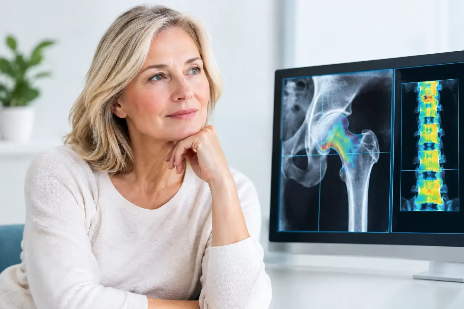
REMS, or Radiofrequency Echographic Multi Spectrometry, uses radiation-free ultrasound to assess bone mineral density at the lumbar spine and proximal femur. It also analyzes the raw ultrasound signals reflected from bone to derive an independent Fragility Score.
The Fragility Score is not another name for density, and it is not a direct measurement of bone flexibility, microarchitecture, or failure load. It reflects signal characteristics associated with skeletal fragility that density alone does not capture. REMS then considers density and the Fragility Score together to estimate five-year fracture risk.
That creates a more complete set of questions for someone going through menopause:
- How much mineralized bone is present?
- Is the bone showing ultrasound signal features associated with greater fragility?
- Taken together, what do those findings suggest about fracture risk over the next five years?
For a person with a lower density result, the added fragility information can help clarify whether both measures point in the same direction or tell a more nuanced story. For a person whose density seems reassuring, it may reveal why density should not be treated as the only relevant number. Neither scenario replaces clinical judgment, but both can make a clinician conversation more specific.
REMS is FDA-cleared and does not use ionizing radiation. Its precision also matters when monitoring is the goal. Small changes in bone measures can be difficult to distinguish from ordinary measurement variation. With an intra-operator precision error below 0.5%, REMS can support repeat measurement over shorter intervals when clinically appropriate, rather than treating bone as something that can only be revisited years later.
What you can control, and what you can clarify
Menopause is one influence on bone, not the whole explanation. Peak bone mass is largely established by early adulthood, and genetics strongly influences the ceiling each person starts with. That is not a reason for resignation. It is a reason to understand your own baseline rather than borrowing reassurance or concern from someone else’s results.
Mechanical loading gives bone a stimulus to adapt. Adequate nutrition, including nutrients involved in bone metabolism such as calcium and vitamin D, supports the biological processes involved in maintaining bone. Sleep, smoking status, alcohol exposure, medications, endocrine health, and fall risk also matter. These are not interchangeable levers, and their relevance depends on your health history.
If a bone result raises questions, the next step is interpretation in context. A report can be brought to a primary care clinician, gynecologist, endocrinologist, or other qualified provider alongside information about menstrual history, medications, fractures, family history, and other conditions. The value is not in collecting another number for its own sake. It is in making a decision with a clearer picture of what the number means.
For people in Western Washington or the Phoenix area, MobileREMS provides access to this radiation-free assessment outside the narrow moment when a fracture or diagnosis finally demands attention.
Menopause changes more than a calendar. It can change the pace of bone remodeling while nothing hurts and nothing looks different. Knowing your density is worthwhile. Knowing whether the signal from your bones adds evidence of fragility, and what both measures suggest about fracture risk, gives you a more useful place to begin.