
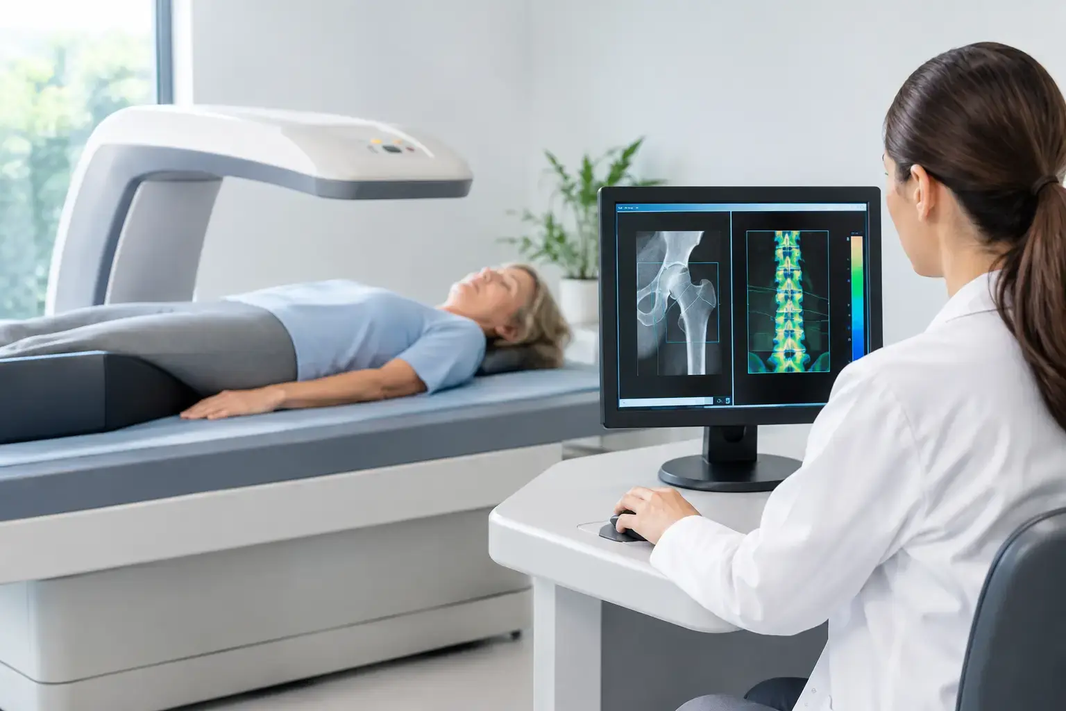
A bone baseline is most useful before a fracture, a troubling scan result, or a major health transition forces the question. The best bone baseline tests do more than assign a single density number. They establish how much mineralized bone is present, look for signs that fracture risk may be higher than density suggests, and identify medical factors that can affect the skeleton.
That distinction matters because bone loss is quiet. You can track blood pressure, glucose, strength, and body composition for years while knowing very little about the condition of the structure carrying you through all of it.
What a meaningful bone baseline needs to answer
There is no one test that is best for every person. A useful baseline is a set of information chosen for the question in front of you: prevention, menopause transition, osteopenia, type 2 diabetes, medication decisions, rapid weight loss, or preparation for orthopedic care.
At minimum, the assessment needs to separate three related but different questions. How much mineralized bone is present? Is there evidence that the bone is showing greater fragility than density alone reveals? And, when density and fragility information are considered alongside your health history, what might that mean for fracture risk?
A test can be technically accurate and still leave an important question unanswered. Bone mineral density is central to bone health assessment, but it is not a direct measurement of every factor involved in fracture. That is why the strongest baseline is often layered rather than limited to one result.
Bone density testing: the essential starting point
Dual-energy X-ray absorptiometry, usually called DXA or DEXA, remains the standard bone density test used in many diagnostic and treatment pathways. It commonly measures the lumbar spine and hip, sites where density is clinically meaningful and where fractures carry substantial consequences.
DXA reports bone mineral density and compares it with reference populations. For postmenopausal women and men age 50 and older, the T-score is commonly used to describe how a result compares with the average bone density of a healthy young adult. A Z-score compares results with people of a similar age and sex and can be more relevant in other clinical contexts.
Those numbers are valuable. They can identify low bone mass, support an osteoporosis diagnosis in the appropriate setting, and create a reference point for future comparison. But density answers a specific question: how much mineralized bone is present in the measured area. It does not, on its own, fully explain why two people with similar density can have different fracture experiences.
A baseline DXA is particularly useful when you and your clinician need a result that fits established diagnostic frameworks. Its trade-off is that small changes can be difficult to distinguish from normal measurement variation, especially over short intervals. The precision of the specific facility and the consistency of follow-up technique matter when interpreting change.
When a normal density result is not the whole story
Type 2 diabetes is the clearest example of why density alone can be falsely reassuring. People with type 2 diabetes may have normal or even higher bone mineral density while still experiencing higher fracture risk. Diabetes can affect bone turnover and collagen-related bone quality, while neuropathy, vision changes, and certain medications can also influence fall risk. A density number may therefore look reassuring without capturing the full risk picture.
The same principle can matter with long-term glucocorticoid exposure, cancer-treatment histories, rapid weight loss, and other conditions that influence bone biology or fracture risk. This does not mean density testing has failed. It means the result needs context.
REMS: density plus information about fragility
Radiofrequency echographic multi-spectrometry, or REMS, measures bone mineral density at the lumbar spine and proximal femur using radiation-free ultrasound. It also analyzes the raw ultrasound signals reflected from bone to derive an independent Fragility Score.
The distinction is worth making carefully. The Fragility Score is not another version of bone density, and it is not a direct measurement of bone flexibility, microarchitecture, or failure load. It reflects ultrasound-signal patterns associated with skeletal fragility that density alone does not capture.
REMS then considers density and the Fragility Score together to estimate five-year fracture risk. Instead of leaving with one number, you leave with density, a Fragility Score, and an estimated near-term fracture-risk result that can be followed over time.
Picture two people with comparable hip density. One has a lower Fragility Score and the other has a higher one. Their density result may look similar, but the reflected ultrasound data tell a different story about fragility-related signals. That difference is precisely the information density cannot provide by itself.
For someone with osteopenia, that added layer can clarify whether density and fragility are telling a consistent story. For someone with type 2 diabetes or another density-misses-it risk factor, it can add information where a density result alone may feel incomplete.
REMS is FDA-cleared and radiation-free. Its reported intra-operator precision error is below 0.5%, compared with the roughly 0.9% to 1.9% often reported for DXA at the spine. Precision is not a technical footnote. Lower measurement error can make it more practical to reassess over months rather than years, with greater confidence that a change on the report reflects a meaningful biological change rather than instrument noise. MobileREMS uses this technology to bring that information to people and clinical partners in the Seattle and Phoenix areas.
Laboratory testing: looking for contributors, not measuring bone strength
Blood and urine tests do not measure bone density or produce a Fragility Score. Their role is different: they can help identify conditions that contribute to bone loss or alter mineral metabolism.
The appropriate laboratory evaluation depends on your age, history, medications, symptoms, and scan findings. A clinician may consider calcium, kidney function, alkaline phosphatase, 25-hydroxyvitamin D, parathyroid hormone, thyroid testing, complete blood count, or other studies when the history points in that direction. In selected situations, testing for celiac disease, sex-hormone status, or excess cortisol may be relevant.
These tests are not a routine checklist for every person with a bone-health question. They are a way to investigate the biology around the skeleton when there is reason to do so. Vitamin D, for example, participates in calcium absorption and bone metabolism, but a vitamin D result does not tell you your bone density or your fracture risk.
Vertebral fracture assessment and imaging when symptoms or history warrant it
A prior fracture changes the conversation. Vertebral compression fractures can occur with little or no dramatic injury and may be discovered after height loss, persistent back pain, a change in posture, or imaging done for another reason.
Vertebral fracture assessment, often performed with a DXA system, or conventional spine imaging can identify fractures that a density test cannot. This matters because an existing fragility fracture is clinical evidence of skeletal vulnerability, regardless of whether a density result falls above or below a particular threshold.
Imaging is not automatically part of every preventive baseline. It becomes more relevant when symptoms, loss of height, long-term steroid exposure, or a fracture history raises the possibility that an unnoticed vertebral fracture is already part of the picture.
Clinical risk review: the test that happens in conversation
No scan knows whether you have fallen before, used glucocorticoids for years, entered menopause early, lost weight quickly, had a parent with a hip fracture, or started a medication that may affect bone. Those details belong in the baseline too.
A clinical review places test results in context. It can also determine whether a fracture-risk calculator is appropriate. Many commonly used calculators estimate longer-term risk and rely heavily on clinical factors and density, when available. A REMS estimate is different: it incorporates REMS density and Fragility Score to estimate risk over the next five years.
Neither approach turns a person into a prediction. Risk estimates are tools for clearer conversations and better-informed decisions, not verdicts about what will happen to you.
Choosing the best baseline for your situation
If your question is simply whether you have low bone density within standard diagnostic frameworks, DXA may be the central test. If you want density plus an independent measure of fragility-related signals and a five-year fracture-risk estimate, REMS adds information that density alone cannot provide. If your history suggests a potential underlying cause of bone loss, laboratory testing may belong alongside imaging. If there has been height loss, back pain, or a suspected prior fracture, vertebral imaging may be the missing piece.
The point is not to collect every available result. It is to establish a baseline that matches the decision you are trying to make and gives future measurements a meaningful reference.
Your bones do not need to become an emergency before they deserve attention. A well-chosen baseline makes an invisible system more understandable, leaving you with information you can bring to your clinician, revisit over time, and use before consequences have to explain the story for you.