
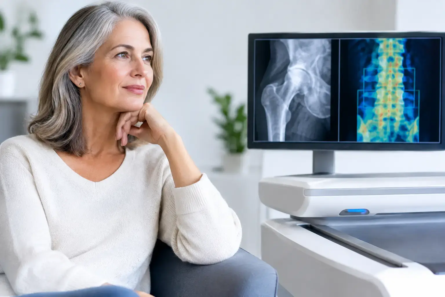
The first years around menopause can be a quiet turning point for your skeleton. You may feel well, remain active, and have no reason to suspect change - yet bone remodeling can shift quickly. So, do bones weaken after menopause? They can. The loss of estrogen accelerates bone loss for many women, particularly through the menopause transition and early postmenopausal years.
That does not mean a fracture is inevitable, or that every person loses bone at the same rate. It means menopause is a biologically meaningful time to understand what is happening beneath the surface, rather than waiting for a fall or a routine screening milestone to begin the conversation.
Why estrogen changes bone remodeling
Bone is living tissue. Throughout adulthood, specialized cells continuously remove older bone and replace it with new bone. This process, called remodeling, repairs small areas of wear and helps maintain the skeleton's mineralized structure.
Estrogen helps regulate that balance. As ovarian estrogen production declines during perimenopause and menopause, the cells that break down bone, called osteoclasts, tend to become more active and live longer. Bone formation does not always keep pace. The result is a net loss of mineralized bone, often most rapidly around the final menstrual period and in the years that follow.
The spine and hip matter especially because they are common sites of osteoporotic fracture and because loss in these areas has real consequences for independence and mobility. But the pace and extent of change vary. Genetics influences the peak bone mass you built earlier in life. Body weight, nutrition, physical activity, smoking, alcohol exposure, certain medications, medical conditions, and prior fractures can all shape what happens next.
Menopause is therefore not one risk factor among many. It is a change in the hormonal environment that can reveal how much skeletal reserve you have - and how your bones are responding now.
Bone density matters, but it is not the whole answer
Bone mineral density, or BMD, measures how much mineralized bone is present in a scanned area. It is useful information and remains central to osteoporosis assessment. A T-score compares your density with the average peak bone density of a healthy young adult reference population. That number can identify low bone mass and osteoporosis and can help track change over time.
But density is not identical to strength, and it is not a complete description of fragility. Two people can have similar bone density measurements while carrying different fracture risk. One may have signal characteristics associated with greater fragility that are not visible in a density number alone. Another may have a density result that looks reassuring while other clinical factors complicate the picture.
Type 2 diabetes is a clear example of the density gap. People with type 2 diabetes can have normal or even higher BMD while experiencing elevated fracture risk. The reasons are still being studied, but altered bone material properties, accumulated glycation-related changes, falls, and complications such as neuropathy may contribute. The point is not that density is unhelpful. It is that a normal density result does not always settle the question of fracture risk.
The same broader question can arise with long-term glucocorticoid use, cancer-treatment histories, rapid weight loss, and other circumstances that affect the skeleton. Density tells you how much mineralized bone is present. A complete conversation also asks whether the bone is showing signs associated with fragility and what the combined information suggests about fracture risk.
Do bones weaken after menopause even with a normal test?
Possibly. A normal or near-normal density result can be reassuring, but its meaning depends on the full context. Bone loss after menopause is not uniform, and a single measurement is a snapshot rather than a lifelong verdict.
Consider two women in their early sixties with similar lumbar spine density. Their density scores may place them in the same category, yet their underlying ultrasound signals may differ in ways associated with fragility. If one also has a prior low-trauma fracture, early menopause, diabetes, or long-term steroid exposure, the clinical conversation becomes more nuanced still.
This is why the question is larger than, “Is my T-score normal?” A more useful set of questions is: How much mineralized bone do I have? Is there independent information suggesting greater fragility? And what does the combination mean for my near-term fracture risk?
No scan replaces clinical judgment. Fracture risk also reflects age, fall history, medications, health conditions, and prior fractures. Still, better skeletal information can make that judgment more informed.
What REMS adds beyond bone mineral density
Radiofrequency Echographic Multi Spectrometry, or REMS, uses radiation-free ultrasound to measure bone mineral density at the lumbar spine and proximal femur. It also analyzes the raw ultrasound signals reflected from bone to derive an independent Fragility Score.
The Fragility Score is not another version of the T-score. It reflects signal features associated with skeletal fragility that density alone does not capture. REMS then weighs density and Fragility Score together to estimate five-year fracture risk.
That creates three distinct pieces of information you can understand and follow over time: bone mineral density, Fragility Score, and estimated five-year fracture risk. Each answers a different part of the same question. Density describes the amount of mineralized bone. The Fragility Score adds information about signs associated with fragility. The risk estimate puts the two together in a forward-looking frame.
This distinction matters during and after menopause because the biological change may begin well before a person reaches the age when routine screening is commonly discussed. A baseline does not predict every future outcome, but it can turn an invisible process into something observable. Follow-up measurements can then show whether the numbers are stable or changing.
REMS is FDA-cleared and radiation-free. Its reported intra-operator precision error is below 0.5%, compared with the approximately 0.9% to 1.9% typically reported for DXA at the spine. Precision matters because small changes can otherwise be difficult to separate from measurement noise. When a test is precise, repeat assessment over months can provide more confidence that a change reflects the bone rather than the instrument.
What can influence bone health after menopause
The menopause transition is central, but it does not act alone. Mechanical loading stimulates bone adaptation because bone tissue responds to repeated forces. Adequate protein, calcium, and vitamin D support different aspects of bone metabolism, with vitamin D helping the body absorb calcium. Sleep, smoking, alcohol, body weight changes, medications, and medical conditions also influence the picture.
This is where broad advice can become misleading. Weight-bearing activity may support bone health, for example, but an appropriate approach differs for someone with a recent fracture, severe osteoporosis, joint disease, cancer treatment, or a high level of athletic training. Medication decisions similarly depend on fracture history, density, age, risk factors, and individual health circumstances. Bone data can support those conversations; it does not substitute for them.
When menopause is a reason to look closer
You do not need symptoms to have changing bones. Osteoporosis is often silent until a fracture occurs, which is precisely why early information has value. Menopause may be an especially useful time to establish a baseline if you have experienced early menopause, a family history of hip fracture or osteoporosis, a previous adult fracture from a low-impact event, substantial weight loss, or exposure to medications known to affect bone.
It can also be useful if you are already tracking cardiovascular fitness, glucose, body composition, or other health measures but have never looked beyond a bone density number. Your skeleton is not separate from the rest of your health. It is a dynamic tissue responding to hormones, metabolism, activity, and time.
A menopause-related bone assessment is not about predicting disaster. It is about replacing assumption with information: how much bone is present, whether there are signs associated with fragility, and what those findings may mean together. That is a calmer, more useful place to begin than waiting for a consequence to make the question urgent.