
A normal bone-density result can be reassuring, but it may not answer the question you actually need answered: how vulnerable are your bones to fracture? If you are asking, “does ultrasound measure bone quality,” the most accurate answer is yes, but with an important distinction. Ultrasound-based REMS assesses information related to bone fragility beyond bone mineral density. It does not turn the broad clinical idea of “bone quality” into one simple, complete measurement.
What people mean by bone quality
Bone mineral density, or BMD, measures how much mineralized bone is present at a site such as the lumbar spine or hip. It is a valuable measurement and remains central to bone-health assessment. But density is not the whole explanation for why one person fractures and another person with a similar density does not.
“Bone quality” is a broad term used to describe characteristics that influence skeletal strength and fragility beyond the quantity of mineralized bone. Depending on the context, the term may refer to the organization of bone tissue, changes associated with aging or disease, the material properties of bone, and other factors that affect how bone responds to force.
That breadth matters. No test should be described as a direct, complete inventory of every component of bone quality. A scan may provide meaningful information about fragility without directly measuring every microscopic feature or predicting exactly how a bone would behave in a particular fall.
The practical question is more useful: can a test identify signs that your bones may be more fragile than density alone suggests? With REMS, the answer is yes.
Does ultrasound measure bone quality directly?
Standard diagnostic ultrasound and ultrasound-based bone assessment are not the same thing. An ultrasound image of a tendon, for example, is not a bone-fragility assessment. The method and the signal analysis matter.
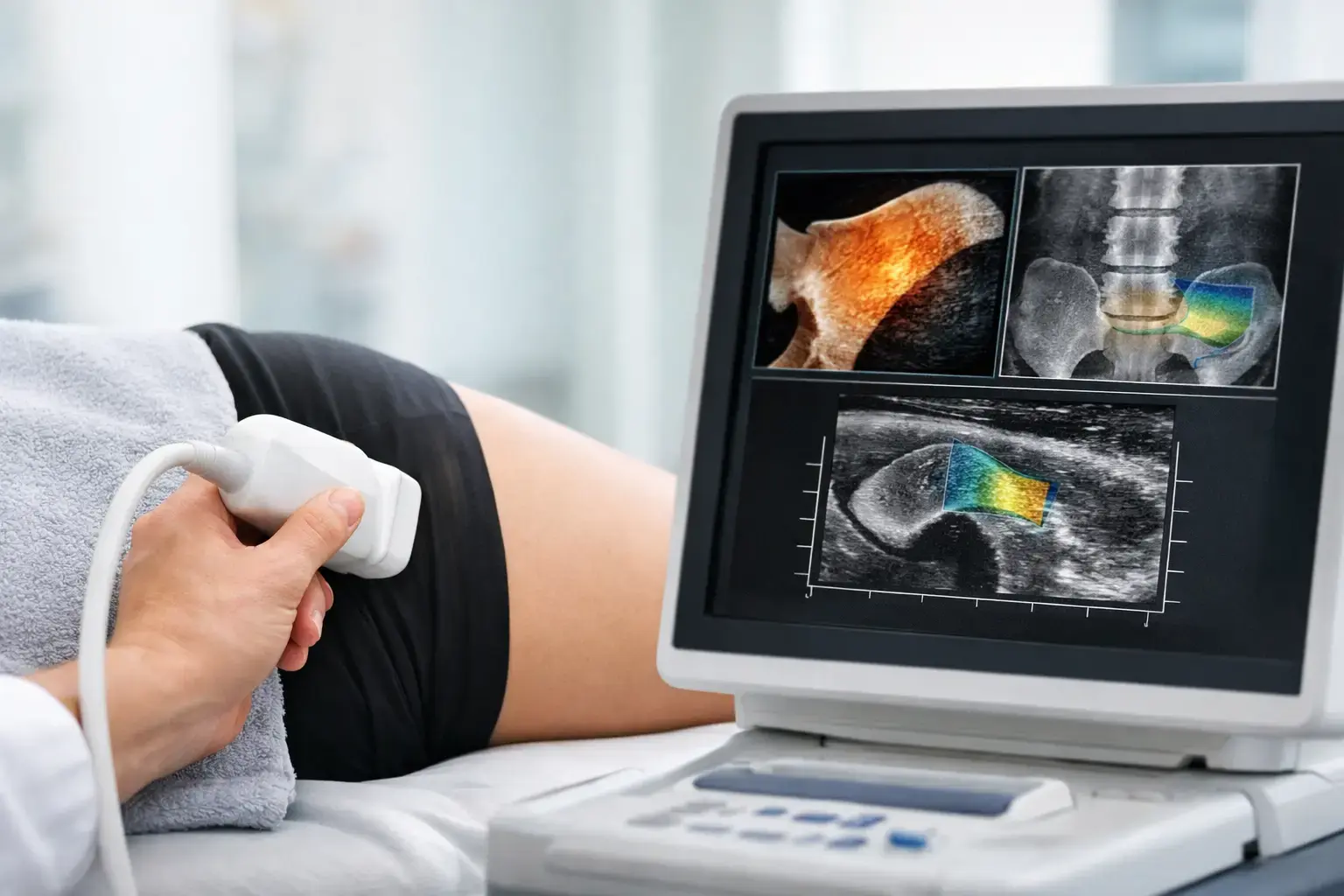
REMS, which stands for Radiofrequency Echographic Multi Spectrometry, uses radiation-free ultrasound at the lumbar spine and proximal femur. It measures bone mineral density at those clinically important skeletal sites. Then it analyzes the raw ultrasound signals reflected from bone to derive an independent Fragility Score.
The Fragility Score is not simply another way of stating your density result. It reflects patterns in the reflected ultrasound signal associated with greater skeletal fragility. REMS combines BMD and the Fragility Score to estimate five-year fracture risk.
That creates a more complete conversation than density alone can provide. You can ask three separate questions:
- How much mineralized bone is present?
- Is the reflected ultrasound signal showing signs associated with greater fragility?
- What do those two measures suggest about estimated fracture risk over the next five years?
These are related questions, but they are not interchangeable. A T-score tells you where your bone density falls compared with a reference population. A Fragility Score adds separate information from the ultrasound signal. Fracture-risk estimation brings those measurements together into a forward-looking clinical context.
Why density and fragility can tell different stories
Imagine two people whose hip-density results fall in the same range. On paper, they appear to have similar amounts of mineralized bone. Yet their reflected ultrasound signals may differ, producing different Fragility Scores and different estimated fracture risks.
This is not a contradiction. It is the gap between bone quantity and the broader question of fragility.
Type 2 diabetes illustrates the gap particularly well. People with type 2 diabetes can have normal, and sometimes higher, bone mineral density while still experiencing elevated fracture risk. One proposed mechanism involves the long-term effects of elevated glucose on bone collagen, including the formation of advanced glycation end products. Bone density may remain reassuring while changes affecting fragility are not fully captured by that density number.
The same need for more information can arise with long-term glucocorticoid exposure, cancer-treatment history, rapid weight loss, and periods of hormonal transition. During perimenopause and menopause, bone loss can accelerate while routine screening may still feel distant. A density test remains useful, but it may leave a thoughtful person asking whether the result tells the entire story.
It does not have to be an either-or choice between density and fragility information. Density matters. The point is that density alone has limits, especially when your health history or clinical picture suggests risk that a single number may not fully explain.
What REMS adds to a bone-density assessment
A REMS examination provides BMD, an independent Fragility Score, and an estimated five-year fracture risk. Each result answers a different part of the bone-health question.
BMD remains the familiar foundation. It shows how much mineralized bone is present and supports established classifications such as osteopenia and osteoporosis. The Fragility Score adds signal-derived information associated with skeletal fragility that density does not capture on its own. The five-year fracture-risk estimate translates the two measures into a result that can be easier to discuss with a physician.
This distinction can be especially meaningful if you have already been told that you have osteopenia or osteoporosis. A diagnosis may give you a T-score but still leave uncertainty: Is my fragility information consistent with that score? Is my risk changing over time? Is a reassuring density result enough in light of my other health factors?
A REMS report cannot answer every clinical question, and it does not replace medical judgment. Fracture risk also involves age, prior fractures, medications, fall history, health conditions, and other variables. But it can add objective information that helps make a conversation more specific.
Why radiation-free precision matters for monitoring
The radiation-free nature of REMS is significant, particularly for people who want to understand change over time. It also measures with high reported intra-operator precision error, below 0.5%. For comparison, DXA precision error at the spine is commonly reported in the range of 0.9% to 1.9%.
That difference is more than a technical detail. Every measurement contains some degree of variation. If a test has more measurement noise, a small change may be difficult to separate from normal test-to-test variation. Tighter precision makes it more feasible to re-measure over months rather than years and have greater confidence that a meaningful change on the report reflects a change in the bone measurement, not simply the instrument.
Monitoring does not mean chasing small fluctuations or interpreting a scan in isolation. It means having data that can be compared thoughtfully in the context of your health, medication decisions, nutrition, activity, hormonal status, and medical history. Mechanical loading stimulates bone adaptation, and vitamin D plays a role in bone metabolism, but how those facts apply to an individual belongs in a conversation with a qualified clinician.
What an ultrasound bone scan cannot tell you
Clear information includes clear boundaries. REMS does not directly measure bone flexibility, failure load, or every aspect of microscopic bone architecture. It does not diagnose the cause of low bone density, identify every condition that affects fracture risk, or guarantee whether you will or will not fracture.
It also should not be treated as a reason to dismiss a DXA result, a prior fracture, or a physician’s evaluation. DXA has decades of clinical use and remains an important tool. The value of REMS is not that it makes density irrelevant. Its value is that it adds independent fragility information to the density picture, without ionizing radiation.
For someone who has never assessed bone health, that may mean establishing a more informative baseline. For someone living with osteopenia, osteoporosis, or a condition such as type 2 diabetes, it may mean moving beyond the question, “What is my T-score?” to ask, “What does the available evidence say about my fragility and near-term fracture risk?”
Bones are often silent until they are not. The goal of better assessment is not to create fear around that silence. It is to make the invisible understandable early enough for you and your clinical team to have a more informed conversation.