
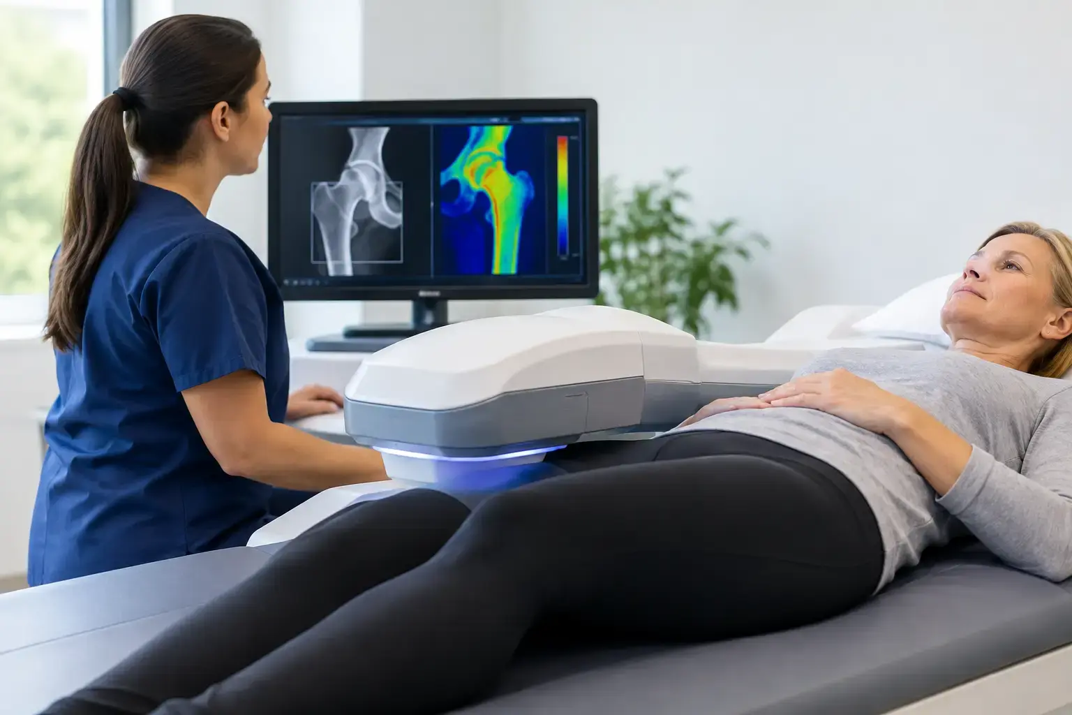
If you are wondering how to prepare for a REMS scan, the answer is reassuringly simple: you generally do not need to fast, change your routine, or prepare for radiation exposure. The more meaningful preparation is knowing what question you want the scan to answer. For many people, that question is larger than, “What is my bone density?” It is, “What does the information in my bones suggest about fragility and fracture risk?”
A REMS assessment measures bone mineral density at the lumbar spine and proximal femur using radiation-free ultrasound. It also analyzes the raw ultrasound signals reflected from bone to derive an independent Fragility Score, then considers both measures in an estimated five-year fracture risk. Preparing well means arriving ready to understand those three pieces of information together.
What you do and do not need to do before a REMS scan
REMS does not use ionizing radiation, so there is no radiation dose to manage before or after the appointment. In most cases, there is no fasting requirement or special dietary preparation. Continue normal daily activities unless the clinic providing the scan gives you different instructions for your appointment.
Medication changes belong with the clinician who manages your care. A bone assessment appointment is not, by itself, a reason to stop or alter a prescribed medication, supplement, or hormone therapy. If you have questions about whether a treatment could affect your results, bring that question to the appointment and discuss the report with your physician or qualified clinician afterward.
Clothing is the practical detail worth confirming in advance. REMS measures the lumbar spine and upper femur, so the sonographer needs access to the lower back or abdominal area and the hip region. Comfortable, two-piece clothing can make this easier. Your scan provider can tell you exactly what will be needed and whether you may need to change clothing for the assessment.
Bring the context that gives your scan meaning
A scan produces measurements. Your health history helps place those measurements in context.
If you have previous DXA reports, prior REMS reports, imaging reports, or laboratory results related to bone health, bringing copies can be useful. So can having a current medication list and a clear account of relevant history, such as menopause status, a prior low-trauma fracture, long-term glucocorticoid use, diabetes, cancer treatment, rapid weight loss, or orthopedic procedures.
You do not need a perfect file folder to have a useful scan. But a few details can sharpen the conversation afterward. A prior T-score, for example, tells you where density was measured at a particular time. A Fragility Score and estimated five-year fracture risk add different information. Seeing how those numbers align, or do not align, can make the next clinical conversation more specific.
It also helps to arrive with one or two questions written down. You may want to know whether your density and Fragility Score point in the same direction, whether the result establishes a meaningful baseline, or what changes would be large enough to follow over time. A report is more valuable when it answers the decision you are actually facing.
What happens during the appointment
A REMS scan is an ultrasound-based assessment, not an X-ray. The operator uses an ultrasound probe at the lumbar spine and proximal femur, the upper part of the thigh bone near the hip. Gel is applied to help transmit the ultrasound signal, much as it is in other ultrasound examinations.
The device measures bone mineral density at those sites. It then evaluates characteristics of the reflected ultrasound signal to generate the Fragility Score. This is the key distinction: REMS is not simply another way to produce a density number. The signal analysis provides information associated with skeletal fragility that density alone does not capture.
The assessment is noninvasive and radiation-free. You will be positioned as needed for the spine and hip measurements, and the operator will guide you through each step. If a position is uncomfortable because of pain, limited mobility, or a recent procedure, say so. The goal is a technically sound measurement without asking you to push through unnecessary discomfort.
Why the Fragility Score changes the conversation
Bone mineral density remains useful. It tells you how much mineralized bone is present. But density does not tell the entire story of fracture risk.
Consider two people with similar density at the hip. On paper, their T-scores may look nearly identical. Yet one person’s reflected ultrasound signal may carry more signs associated with fragility than the other’s. Their independent Fragility Scores may differ, and therefore their estimated five-year fracture risks may differ as well. That is not a contradiction in the data. It is the reason density alone can be incomplete.
Type 2 diabetes makes this information gap especially clear. People with type 2 diabetes can have bone density that appears normal, or even higher than expected, while fracture risk remains elevated. One proposed biological explanation involves chronic hyperglycemia and advanced glycation end products in bone collagen, which may affect skeletal tissue in ways a density measurement does not show. REMS does not measure advanced glycation end products directly. Its value is that it adds an independent ultrasound-derived Fragility Score rather than asking density to answer every question about fracture risk.
The same principle can matter in other situations where the clinical picture is more complicated than a single density result: long-term glucocorticoid exposure, cancer-treatment history, rapid weight loss, or the menopause transition. A scan cannot diagnose every cause of skeletal risk. It can make the invisible information more visible and give you objective results to bring into a broader clinical evaluation.
How to prepare for a REMS scan if you already have a diagnosis
If you have been told you have osteopenia or osteoporosis, you do not need to arrive expecting the scan to either confirm or overturn everything you have heard. Think of it as a way to add resolution.
Your density result remains part of the picture. The Fragility Score is a separate measure derived from the ultrasound signal, and the five-year fracture-risk estimate weighs both. Sometimes all three results tell a consistent story. Sometimes density looks less concerning than the fragility-related information suggests, or the reverse. Either result is useful because it gives your clinician a more complete basis for discussion.
This is also why prior reports matter. A single measurement is a starting point. Follow-up measurements can show whether a result is stable or changing. REMS has an intra-operator precision error below 0.5%, compared with the 0.9% to 1.9% commonly reported for DXA at the spine. That lower measurement error matters because it can make smaller changes more interpretable. It does not mean every small difference is automatically biologically meaningful, but it reduces the chance that apparent change is merely instrument noise.
After the scan: read the report as three related answers
When you receive your report, look beyond any single label or number. The useful questions are straightforward: How much mineralized bone is present? Is the ultrasound signal showing greater or lower signs associated with fragility? What does the combination suggest about estimated fracture risk over the next five years?
A result is not a verdict on your bones. It is information about their current state, measured at clinically relevant sites and interpreted through more than density alone. Whether the report becomes a baseline, prompts a conversation about treatment, or simply fills a long-standing information gap depends on your history and the decisions in front of you.
The best preparation for a REMS scan is not anxiety or elaborate routine changes. It is curiosity, context, and a willingness to ask a better question: not only how much bone do I have, but what can my bones tell me before a fracture has to?