
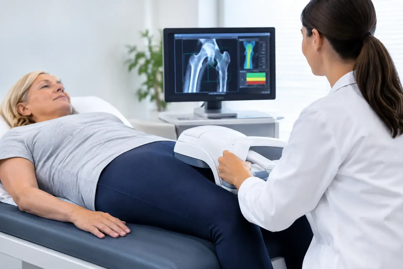
A bone scan can sound more intimidating than it is, especially if you have spent years associating bone testing with X-rays, radiation exposure, or a diagnosis you are not ready to face. REMS scan safety begins with a meaningful distinction: REMS uses ultrasound, not ionizing radiation, to assess bone at the lumbar spine and proximal femur.
That matters if you want a baseline before menopause, are monitoring known osteopenia or osteoporosis, or are living with a risk factor that density may not fully explain. But radiation-free does not mean a scan is only a technical curiosity. It means you can consider bone information without adding radiation exposure to the decision.
What happens during a REMS scan?
REMS, short for Radiofrequency Echographic Multi Spectrometry, is an FDA-cleared ultrasound technology for bone assessment. During the exam, an ultrasound probe is used over the lower back and hip region to acquire signals reflected from bone. There are no injections, contrast agents, or enclosed imaging equipment.
The scan assesses bone mineral density at the same clinically important regions commonly measured by DXA: the lumbar spine and proximal femur. It then analyzes the raw ultrasound signals rather than stopping at density alone. From those signals, REMS derives an independent Fragility Score and combines that information with density to estimate five-year fracture risk.
For most people, the physical experience is straightforward. You are positioned for the measurement while a trained operator acquires images at the spine and hip. The operator may apply gentle pressure with the probe to obtain a clear signal. If a position is uncomfortable because of recent surgery, back pain, hip pain, or limited mobility, say so before the exam begins. Positioning can often be adapted within the limits of obtaining an interpretable measurement.
Why radiation-free is the central safety question
DXA uses a very low dose of ionizing radiation. It has an established role in bone care and remains widely used. REMS is different because ultrasound uses sound waves rather than X-rays to obtain its measurements.
Ionizing radiation has enough energy to remove electrons from atoms. In medical imaging, exposure is managed carefully and the dose from DXA is low, but it is still a form of radiation. Ultrasound works through reflected sound waves. That is why a REMS scan does not add ionizing radiation exposure.
This difference has practical implications for repeat measurement. Bone is living tissue, and its changes may matter over months, not only years. A radiation-free method removes one consideration from the monitoring conversation. It does not mean every person needs frequent scans, or that a number should be interpreted in isolation. It means radiation exposure is not the limiting factor it can be with X-ray-based imaging.
REMS also has a reported intra-operator precision error below 0.5%, compared with approximately 0.9% to 1.9% typically reported for DXA at the spine. Precision is part of safety in the broader clinical sense: if a measured change is mostly machine noise, it can create unnecessary concern or false reassurance. Tighter precision helps make serial measurements more meaningful when they are clinically appropriate.
Safety is also about getting the right information
A safe test is not simply one that feels comfortable and avoids radiation. It must also answer the question you actually need answered.
Bone mineral density is useful. It tells you how much mineralized bone is present. Yet density does not fully account for skeletal fragility. Consider two people with the same density result: one may have ultrasound signal characteristics associated with greater fragility, while the other may not. Their density number is similar, but the fuller fracture-risk picture may differ.
This gap is especially relevant in type 2 diabetes. People with type 2 diabetes can have bone mineral density that appears normal or even elevated while their fracture risk remains increased. The mechanisms are complex and may include changes in bone material properties related to chronic high blood sugar, as well as falls risk and other factors. The point is not that density is wrong. It is that a reassuring density result may not be the whole story.
REMS adds an independent Fragility Score from reflected ultrasound signals. It does not directly measure bone flexibility, failure load, or every aspect of bone microarchitecture. It provides a distinct signal-based measure of fragility alongside density, then uses both to estimate five-year fracture risk. That added context can be valuable when your question is larger than, “What is my T-score?”
Who may need extra context before scanning?
A REMS scan is noninvasive, but the report still needs to be interpreted in the context of your health history. Share relevant information with the scanning provider, particularly if you have had hip or spinal surgery, hardware near the measurement area, a recent fracture, major changes in mobility, or conditions that affect positioning.
These factors do not automatically mean you cannot be scanned. They may affect whether a target area can be measured reliably or whether another assessment is needed for a particular clinical question. The same is true of body shape, anatomy, and degenerative changes: imaging technologies have technical limits, and a useful report should acknowledge them rather than force certainty where it does not exist.
Pregnancy is another situation where context matters. Because REMS does not use ionizing radiation, its radiation profile differs fundamentally from X-ray-based testing. Still, pregnancy-related bone concerns, symptoms, and any testing decision belong in a conversation with the clinician overseeing prenatal care. Radiation-free is a meaningful feature, not a substitute for individualized clinical judgment.
What a REMS result can and cannot do
A REMS report gives you three pieces of information to follow over time: bone mineral density, Fragility Score, and estimated five-year fracture risk. It can provide a clearer starting point for a conversation with your physician or other qualified clinician, especially if you are evaluating osteoporosis medication, monitoring bone health during rapid weight loss, or trying to understand a prior osteopenia result.
It cannot diagnose every cause of bone pain, determine whether a specific exercise or supplement is right for you, or replace medical evaluation after a fall or suspected fracture. New severe back, hip, or groin pain after an injury deserves prompt clinical attention regardless of a prior bone scan result.
There is also a difference between screening information and a treatment decision. A scan may reveal that density and fragility are telling a consistent story, or that they are not. Either finding can improve the quality of the next conversation. The result is information to bring into care, not a verdict on your bones.
A calmer way to think about bone testing
For many adults, the most reassuring aspect of REMS scan safety is not merely that the exam is radiation-free. It is that the technology can make an invisible process more understandable before a fracture forces the issue.
Your bones are not defined by a single density number, and a bone scan is not a reason to assume the worst. It is a way to see more clearly: how much mineralized bone is present, whether the ultrasound signal shows signs associated with greater fragility, and what those findings may mean together for fracture risk.