
Menopause can change your bones long before anything hurts. The top menopause bone tests are not interchangeable, because they answer different questions: how much mineralized bone you have, whether silent fractures may already be present, and whether another health condition could be contributing to bone loss.
That distinction matters during the menopause transition. Falling estrogen accelerates bone remodeling, the normal cycle in which older bone is removed and new bone is formed. When removal outpaces replacement, bone mineral density can decline quickly. A test that gives only one number can be useful, but it may not be the whole conversation about skeletal fragility.
Start with the question, not the test
A bone assessment is most useful when it matches the decision in front of you. Someone seeking an early baseline has different needs from someone with a prior low-trauma fracture, an osteopenia diagnosis, or a new medication that may affect bone.
The central question is often framed as, “Do I have osteoporosis?” But a more informative set of questions is: How much mineralized bone is present? Is there evidence that my bones are more fragile than density alone suggests? What is my estimated fracture risk? And are there medical factors that need attention?
Here is how the main menopause bone tests fit together.
DXA: the standard bone density test
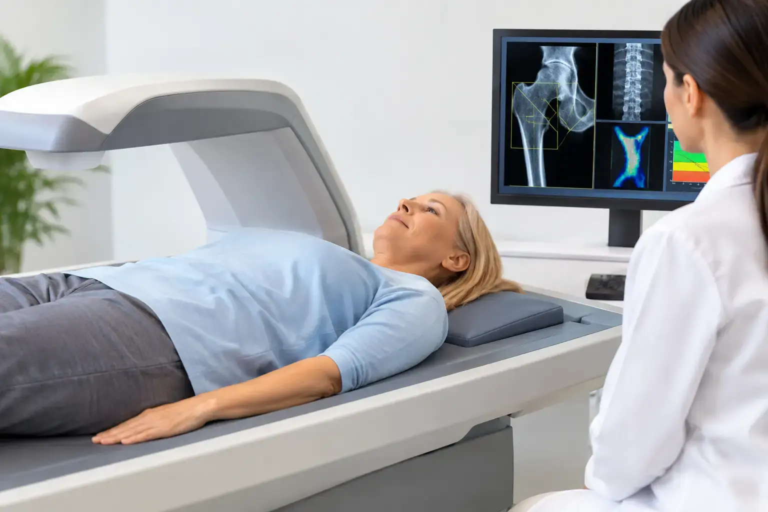
Dual-energy X-ray absorptiometry, usually called DXA or DEXA, remains the standard test for diagnosing osteoporosis. It measures bone mineral density, most often at the lumbar spine and hip. The report compares your result with reference populations and provides T-scores and Z-scores.
A T-score compares your density with the average peak bone density of a healthy young adult. In general, a T-score from -1.0 to -2.5 is categorized as osteopenia, while a score of -2.5 or lower is categorized as osteoporosis. These thresholds are clinically established and useful for diagnosis, treatment discussions, and follow-up.
DXA has limits that matter in menopause. Density is an amount measurement. It tells you how much mineralized bone is present, not every feature associated with skeletal fragility. Two people can have similar T-scores and different fracture outcomes because bone fragility is influenced by more than mineral quantity.
DXA results can also be affected by technical and anatomical factors. Degenerative changes in the spine, arthritis, and calcification can sometimes make spinal density appear higher. That does not make DXA unhelpful. It means the result is best read in context, alongside the hip measurement, fracture history, age, medications, and other risk factors.
For many women, DXA is the right starting point. Routine screening is commonly recommended at age 65, with earlier assessment considered when risk factors are present. Menopause itself is a biological transition worth discussing sooner, particularly after early menopause, surgical removal of the ovaries, a fracture from a low-impact event, prolonged glucocorticoid use, or substantial weight loss.
REMS: density plus information about fragility
Radiofrequency Echographic Multi Spectrometry, or REMS, also measures bone mineral density at the lumbar spine and proximal femur. It uses ultrasound rather than ionizing radiation. But its additional value is in what it reads from the reflected ultrasound signals.
Those raw signals are used to derive an independent Fragility Score, which reflects signal features associated with skeletal fragility that density alone does not capture. REMS then weighs bone mineral density and the Fragility Score together to estimate five-year fracture risk.
This creates a more complete conversation than a density number by itself. Imagine two women whose hip density falls in the same range. One has a lower Fragility Score and a lower estimated fracture risk; the other has a higher Fragility Score despite similar density. The density result has not become irrelevant. It is simply no longer the only information available.
That distinction can be especially meaningful through menopause, when you may want to understand change before a fracture forces the issue. MobileREMS uses FDA-cleared REMS technology to provide three followable measures: density, Fragility Score, and estimated five-year fracture risk.
Precision also affects how often a measurement can be useful. REMS has an intra-operator precision error below 0.5%, compared with the approximately 0.9% to 1.9% commonly reported for DXA at the spine. Lower measurement error does not predict your future by itself. It does make it easier to distinguish a meaningful change in bone measurements from ordinary testing noise when monitoring is clinically appropriate.
REMS does not replace every role of DXA, and availability, insurance coverage, prior testing, and your clinician’s needs all matter. It adds information beyond density, rather than turning bone health into a single perfect score.
Vertebral fracture assessment: looking for fractures you may not feel
Vertebral compression fractures are common in osteoporosis and can occur with little or no memorable injury. Some are discovered only after height loss, a change in posture, or imaging obtained for another reason. Yet a vertebral fracture can substantially change a person’s fracture-risk picture.
Vertebral fracture assessment, often performed with certain DXA systems, uses low-dose imaging to look for fractures in the thoracic and lumbar spine. Standard spine X-rays may also be used when symptoms or clinical findings warrant a closer look.
This is not a substitute for a density test or a fragility assessment. It answers a separate, highly practical question: Is there evidence that a fracture has already occurred? If you have lost height, developed persistent back pain, or have known osteoporosis, a clinician may consider whether vertebral imaging is relevant.
FRAX: a risk calculation, not a scan
FRAX is often discussed alongside menopause bone tests, but it is not an imaging test. It is a fracture-risk calculator that combines clinical factors such as age, prior fracture, smoking, glucocorticoid use, rheumatoid arthritis, and alcohol use. It can be calculated with or without femoral neck bone mineral density.
Its strength is that it moves the conversation beyond a T-score. Its limitation is that any calculator depends on the factors it includes. FRAX does not directly measure the structural signals used to generate a REMS Fragility Score, and it may not fully reflect every source of risk in every person.
Think of it as one lens for organizing established risk factors. A thoughtful bone-health assessment may include FRAX alongside density, fracture history, and other clinical information rather than treating it as a final verdict.
Blood and urine tests: finding contributors to bone loss
Laboratory tests do not measure bone density or tell you whether a bone will fracture. Their role is different: they can help identify conditions that affect bone metabolism or contribute to bone loss.
Depending on your history, a clinician may evaluate vitamin D status, calcium, kidney function, thyroid function, parathyroid hormone, blood count, liver markers, or markers of inflammation and malabsorption. In selected situations, bone turnover markers may help show the pace of bone remodeling, although they are not a stand-alone diagnostic test for osteoporosis.
This is particularly relevant when bone loss seems faster than expected, menopause occurred early, there is a history of digestive disease, or medications may affect bone. The goal is not to order every available lab. It is to avoid assuming that menopause is the only explanation when another contributor may be present.
When density may not tell the whole story
Menopause is not the only reason density and fracture risk can diverge. Type 2 diabetes is the clearest example. People with type 2 diabetes can have normal or even higher bone mineral density while still experiencing elevated fracture risk. Changes in bone material properties, falls risk, and diabetes-related complications may all contribute.
Long-term glucocorticoid exposure, cancer-treatment history, rapid weight loss, and some other medical circumstances can also complicate a density-only interpretation. None of this means density is unimportant. It means a reassuring density result should be interpreted alongside the biology and the person carrying that result.
Choosing among the top menopause bone tests
There is no single best test for every woman in menopause. DXA establishes the standard density measure. REMS adds an independent Fragility Score and a five-year fracture-risk estimate from radiation-free ultrasound. Vertebral imaging can identify silent fractures, while laboratory testing can investigate potential drivers of bone loss. FRAX can bring clinical risk factors into the discussion.
The useful next step is not to chase more numbers for their own sake. It is to ask for information that makes your bone health more understandable: what your density shows, what it cannot show on its own, and whether your current life stage calls for a fuller picture before consequences make the decision for you.