
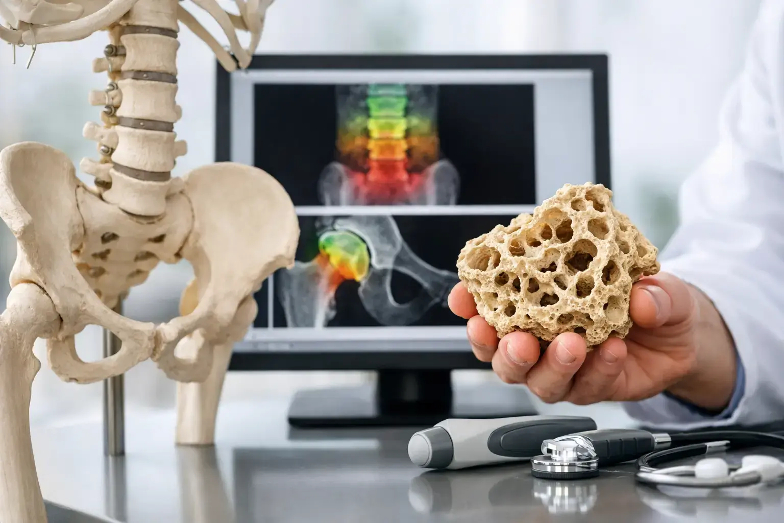
A bone density result can look reassuring and still leave a meaningful question unanswered: how fragile is the bone itself? Skeletal fragility is the part of bone health that becomes visible when a bone does not hold up as expected, often long before a fracture makes the issue impossible to ignore.
That distinction matters whether you are entering menopause, living with osteopenia, managing type 2 diabetes, or simply paying attention to the health data that shape your future. Bone mineral density is useful. It tells you how much mineralized bone is present. But a density number alone cannot fully explain why two people with similar results may have different fracture risk.
Skeletal Fragility Is Not the Same as Low Density
Bones are living tissue, continually responding to hormones, nutrition, mechanical loading, illness, medication exposure, and aging. Density describes one measurable property of that tissue: the amount of mineralized bone in a given area. Lower density is associated with higher fracture risk, which is why density remains central to bone assessment.
But fracture risk is not determined by mineral quantity alone. A useful comparison is two houses built with the same amount of lumber. One may have materials arranged and maintained in a way that better withstands a storm. The other may show signs associated with greater vulnerability, despite having the same total amount of wood. Bones are not houses, and this is not a literal measure of bone structure. It illustrates the question density cannot answer by itself: does the tissue show signal patterns associated with greater fragility?
That is why skeletal fragility deserves its own conversation. It refers to the tendency of bone to fracture under forces that would not ordinarily cause a break, or to be more vulnerable than density alone would suggest. Fragility is a clinical outcome and risk concept, not a single physical trait. It is influenced by density, age, prior fracture, falls, medications, medical conditions, and features of bone that standard density measurement does not directly capture.
The false-normal problem in type 2 diabetes
Type 2 diabetes is one of the clearest examples of why density does not tell the whole story. People with type 2 diabetes can have bone mineral density that is normal or even higher than expected, while their fracture risk remains elevated. Large observational studies and meta-analyses have repeatedly documented this mismatch.
Several mechanisms may contribute. Chronic high blood glucose can promote advanced glycation end products, which alter collagen in bone tissue. Diabetes can also affect bone turnover, vision, nerve function, muscle function, and fall risk. No single mechanism explains every person’s risk, but the pattern is clinically relevant: a normal density result does not automatically mean normal skeletal resilience.
This is not a reason to distrust density. It is a reason to interpret density in context. The same principle can matter with long-term glucocorticoid use, cancer-treatment history, rapid weight loss, and other situations where bone risk may not be neatly summarized by one number.
What Density Can Tell You, and What It Cannot
A DXA scan has long been the standard method for measuring bone mineral density. Its T-score compares your result with the average density of a healthy young adult reference population. That information helps identify low bone mass and osteoporosis and can guide important clinical decisions.
Density is particularly valuable as a baseline and as a trend over time. If density changes meaningfully, that can reveal a shift in skeletal health. Yet a density result does not independently report whether the reflected properties of the bone tissue carry signs associated with fragility. Nor does it turn a measurement into a guaranteed prediction of whether you will or will not fracture.
Fractures are multifactorial. Bone strength matters, but so do the circumstances of a fall, balance, mobility, medications, vision, and medical history. A good assessment does not pretend that one result settles every question. It provides clearer information for the decisions and conversations that follow.
How REMS Adds Information About Skeletal Fragility
Radiofrequency Echographic Multi Spectrometry, or REMS, uses radiation-free ultrasound to measure bone mineral density at the lumbar spine and proximal femur. It also analyzes the raw ultrasound signals reflected from the bone. From those signals, REMS derives an independent Fragility Score.
The Fragility Score is not another density score, and it is not a direct measurement of microarchitecture, flexibility, brittleness, or failure load. It reflects ultrasound signal characteristics associated with skeletal fragility. That distinction is worth preserving because it explains why the score can add information rather than simply repeat the density result.
REMS then weighs bone mineral density and the Fragility Score together to estimate five-year fracture risk. The result is a more complete set of questions and answers:
- How much mineralized bone is present?
- Is the bone showing ultrasound signal patterns associated with greater fragility?
- What do those measures, together, suggest about estimated fracture risk over the next five years?
For someone who has been told they have osteopenia, this can help move the conversation beyond a label. Osteopenia describes a density range. It does not, by itself, tell the full story of fragility or define a person’s future. For someone with type 2 diabetes and apparently normal density, the Fragility Score can add context to a result that might otherwise appear uncomplicated.
Precision Changes What Monitoring Can Mean
A measurement is only useful for tracking change if its normal measurement variation is small enough to distinguish change from noise. This is where precision becomes clinically meaningful, not merely technical.
REMS is FDA-cleared and radiation-free, with reported intra-operator precision error below 0.5%. Reported DXA precision at the spine is commonly in the range of 0.9% to 1.9%, although real-world precision varies by site, equipment, positioning, and operator. A lower precision error can make it more feasible to re-measure over shorter intervals and have greater confidence that a visible difference is not simply instrument variation.
That does not mean every person needs frequent testing. The right timing depends on the reason for monitoring, the pace of a health transition, previous results, and the clinical question being considered. It does mean that when bone change may be happening more quickly, measurement precision affects how useful a follow-up result can be.
This matters during menopause, after major changes in weight, when reviewing the effects of a bone-health strategy with a clinician, or when establishing a baseline before a treatment decision. A baseline is not a verdict. It is a reference point from which future changes can be understood.
Reading Your Results Without Overreading Them
The most useful bone report is one that gives you a clearer starting point for a conversation with your physician or other qualified clinician. Density, Fragility Score, and estimated five-year fracture risk each answer a different part of the problem. None should be interpreted in isolation from your medical history.
A lower density result may align with a higher Fragility Score, but the two do not always move together. That is exactly why the information is valuable. Agreement can reinforce a pattern already suggested by density. A mismatch can identify a reason to look more carefully at risk factors that a density number does not represent.
For organizations and practices, offering this kind of assessment can also change the quality of bone-health conversations. Instead of waiting until a fracture creates urgency, patients and members can bring objective information into a discussion earlier. That is especially relevant for people who are already tracking cardiovascular, metabolic, or body-composition data but have never had a comparable view of their bones.
Bone health is often discussed after something happens. Skeletal fragility asks a better question before then: not only how much bone do you have, but what does the available information suggest about how that bone may hold up? Clearer answers do not create fear. They create a more informed place to begin.