
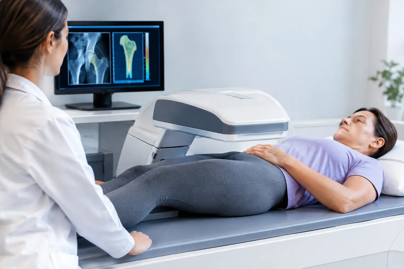
A bone-density result can look reassuring and still leave an unanswered question: what does that number say about whether your bones are showing signs associated with fragility? This is where radiation-free testing options change the conversation. They can provide meaningful bone information without exposing you to ionizing radiation, but the value depends on what the technology actually measures.
For someone navigating menopause, living with osteopenia, managing type 2 diabetes, or simply building a more complete health baseline, the goal is not to replace one number with another. It is to understand what each test can tell you, what it cannot, and whether the result helps answer a real decision in front of you.
Why radiation matters, but information matters more
X-ray-based bone tests use ionizing radiation. In standard bone testing, the exposure from a DXA scan is very low. For most people, that small exposure is not the central limitation of DXA. The more consequential limitation is informational: DXA measures areal bone mineral density, typically at the hip and spine. It tells you how much mineralized bone is present in the scanned area.
That is useful information. It is also not a complete description of skeletal fragility.
A bone's likelihood of fracture is influenced by more than mineral quantity. Clinical factors matter, including age, prior fracture, certain medications, falls, and health conditions. Bone tissue can also show signal patterns associated with fragility that a density value alone does not express. Two people may have similar T-scores and still carry different fracture risk.
Radiation-free testing is appealing because it removes radiation exposure, particularly when repeat measurement is useful. But a test is not automatically more informative because it is radiation-free. The key question is simpler: does it produce accurate, clinically relevant information that adds to what you already know?
The main radiation-free bone testing options
Quantitative ultrasound at peripheral sites
Traditional quantitative ultrasound, often called QUS, is commonly performed at the heel. It uses sound waves rather than X-rays and may measure features such as broadband ultrasound attenuation and speed of sound through the calcaneus.
Heel ultrasound can be convenient and is sometimes used in community screening settings. It may help identify people who warrant a more complete bone evaluation. However, a heel measurement is not the same as a measurement of the lumbar spine or proximal femur, the sites most commonly used to diagnose osteoporosis with DXA. Results from different ultrasound devices are not always interchangeable, and peripheral ultrasound generally does not provide the same type of hip and spine density result used in standard diagnostic thresholds.
Its role is best understood as a screening tool, not a universal substitute for a clinical hip-and-spine assessment.
REMS ultrasound of the spine and hip
Radiofrequency Echographic Multi Spectrometry, or REMS, is a different form of ultrasound assessment. Rather than measuring only at a peripheral site, REMS evaluates the lumbar spine and proximal femur, the same clinically important skeletal regions commonly assessed by DXA.
REMS uses ultrasound to measure bone mineral density without radiation. It also analyzes the raw ultrasound signals reflected from bone to derive an independent Fragility Score. That distinction matters. The Fragility Score is not another label for density; it reflects ultrasound signal characteristics associated with skeletal fragility that density alone does not capture.
The technology then weighs bone mineral density and the Fragility Score together to estimate fracture risk over the next five years. A REMS assessment therefore addresses three related, but distinct, questions: How much mineralized bone is present? Is the bone showing greater signs associated with fragility? What does the combination suggest about near-term fracture risk?
This does not mean an ultrasound scan sees every aspect of bone biology. REMS does not directly measure bone flexibility, failure load, or every feature of microscopic bone structure. Its value is more specific: it adds validated ultrasound-derived information about fragility to a density measurement at the spine and hip.
Other non-ionizing assessments that provide context
Some tools do not measure bone density but can still add context. MRI can examine certain skeletal and soft-tissue questions without ionizing radiation, although it is not a routine replacement for bone-density testing. Laboratory tests can help clinicians investigate contributors to bone loss or altered bone metabolism, such as calcium regulation, vitamin D status, thyroid function, or bone turnover markers.
These tests answer different questions. A blood test may help explain why bone is changing. A bone scan measures the current state of bone at clinically relevant sites. One does not replace the other.
The false-normal problem in type 2 diabetes
Type 2 diabetes is one of the clearest examples of why density alone can be incomplete. People with type 2 diabetes may have bone mineral density that appears normal or even higher than expected, while research consistently shows elevated fracture risk.
The mechanism is still an active area of research, but the mismatch is well recognized. Long-term elevated glucose can contribute to advanced glycation end products in collagen, and diabetes may also affect bone remodeling, muscle function, vision, neuropathy, and fall risk. A density number is measuring mineral quantity. It is not designed to capture all of those pathways.
This does not make density irrelevant for someone with diabetes. It makes context essential. A result that looks normal on one measurement should not automatically end the conversation when other risk information points in a different direction.
The same principle can apply to people using long-term glucocorticoids, those with cancer-treatment histories, people experiencing rapid weight loss, and those considering bone-related medication or orthopedic decisions. The purpose of additional information is not to create alarm. It is to reduce the chance that a single reassuring-looking number is asked to carry more meaning than it can.
Why precision changes the value of follow-up
The practical advantage of radiation-free ultrasound is not only the absence of radiation. It is also the possibility of repeat measurement when timing and precision support a meaningful question.
Every measurement has some degree of variation. If a device's normal measurement error is too large, a small change on a later report may be noise rather than a real biological change. That is why precision is not a technical footnote. It determines how confidently a clinician and patient can interpret change over time.
REMS has an intra-operator precision error below 0.5%, compared with the approximately 0.9% to 1.9% typically reported for DXA at the spine. The practical implication is not that every person needs frequent scanning. Timing depends on the clinical context, the reason for monitoring, and the question being asked. It means the technology is designed to detect smaller changes with less uncertainty when follow-up is appropriate.
For a person who has already received an osteopenia or osteoporosis diagnosis, that can make the report more useful. Instead of watching only a T-score, you can follow density, an independent Fragility Score, and estimated five-year fracture risk. These values do not make decisions for you or your clinician, but they can make the discussion more specific.
How to compare a radiation-free test with DXA
DXA remains an established standard for diagnosing osteoporosis and is widely available. Its radiation dose is low, it has decades of clinical use, and many treatment and insurance pathways are organized around DXA results. A radiation-free test is not inherently a reason to disregard DXA.
The comparison becomes more useful when you consider the information you need. If the question is limited to a conventional T-score for diagnosis or administrative requirements, DXA may be the expected route. If you want hip-and-spine density plus an independent measure of fragility-related ultrasound signals and an estimated five-year fracture risk, REMS provides a broader assessment without radiation.
It also matters that reports can be shared with the clinician who knows your medical history. A scan result belongs alongside fracture history, medications, menopause status, diabetes, mobility, fall history, nutrition, and other relevant health information. No bone test, radiation-free or otherwise, should be interpreted as a complete risk assessment in isolation.
The useful question is not “Which test is best?”
A better question is: what information about your bones is still missing?
If you have never had a baseline, knowing your density at the spine and hip may fill a major gap. If you already know your T-score, the unanswered question may be whether ultrasound signals suggest a different level of fragility than density alone implies. If you are following a change in health, medication, weight, or life stage, precision may matter as much as the initial result.
Your bones are not a future problem waiting for a fracture to make itself visible. They are living tissue, changing quietly over time. The right assessment does not predict every outcome. It gives you and your care team a clearer place to begin the conversation before consequences force it.