
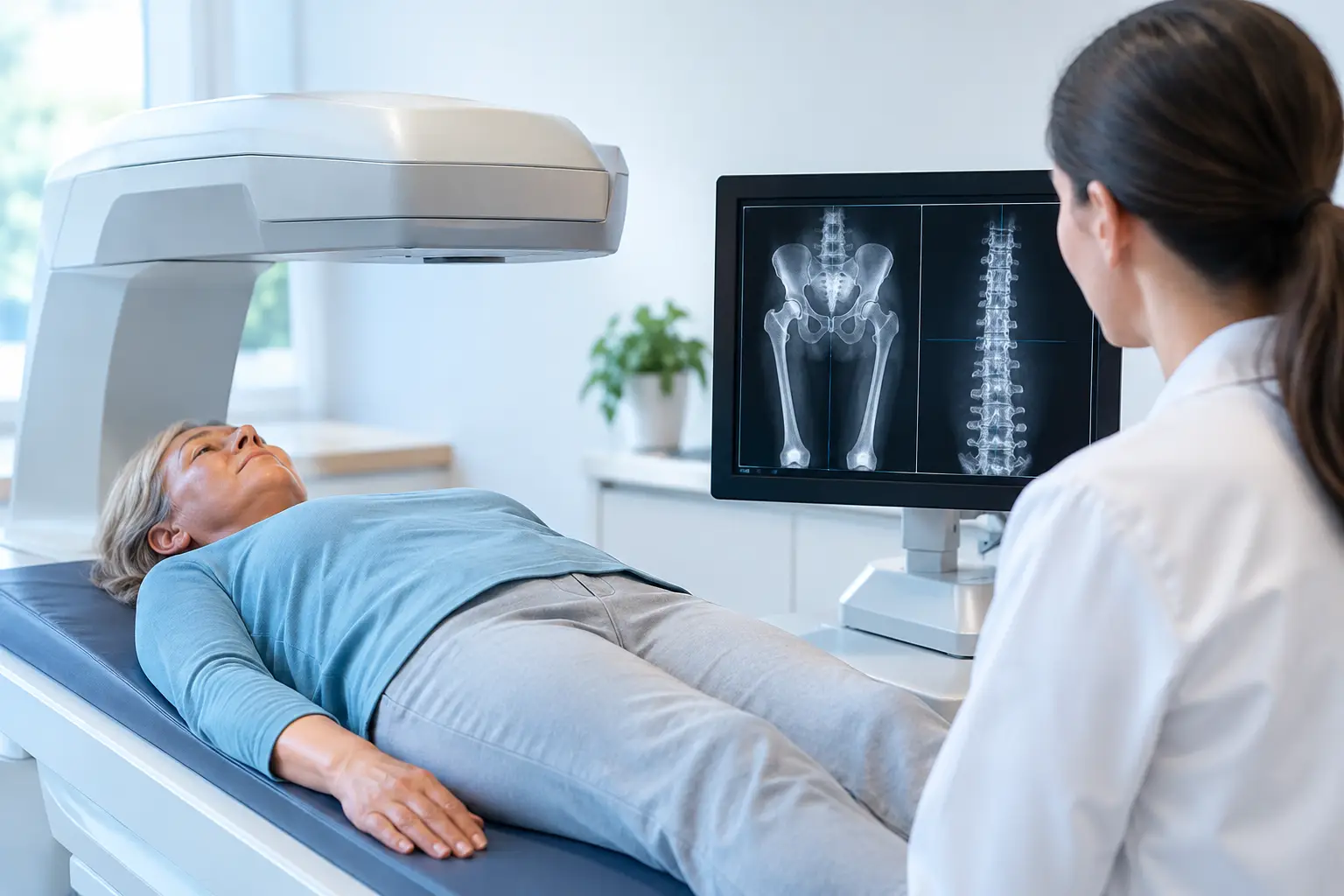
You may be trying to follow a real change in your bones - after menopause, during rapid weight loss, after an osteopenia diagnosis, or while weighing a treatment decision. That makes the question, are frequent bone scans safe, more specific than it first appears: safe compared with what, and frequent enough to answer which question?
The answer depends almost entirely on the scan. “Bone scan” is often used as a catchall term, but a DXA density test, a nuclear medicine bone scan, a CT-based measurement, and a radiation-free REMS assessment are very different examinations. They do not expose you to the same things, measure the same biology, or make sense on the same monitoring schedule.
Are frequent bone scans safe? Start with the type of scan
A conventional DXA scan is the familiar test used to measure bone mineral density at the spine and hip. It uses very low-dose X-rays. The radiation exposure from a standard central DXA exam is generally a small fraction of everyday background radiation and is far lower than that of most CT studies. For most adults, repeated DXA testing is not a meaningful radiation concern.
That does not mean repeating it every few months will necessarily produce useful information. DXA has measurement variability. A small difference between one result and the next can reflect positioning, analysis, equipment, or ordinary test variation rather than a true biological change. This is why the question is not only whether the radiation is safe. It is whether the measurement is precise enough, and the interval long enough, to distinguish change from noise.
A nuclear medicine bone scan is something else entirely. It involves an injected radiotracer that collects in areas of active bone turnover. Clinicians may use it to investigate suspected fractures, certain cancers, infections, or other focal bone conditions. Its radiation exposure is substantially higher than DXA, and it is not a routine test for tracking osteoporosis or gradual bone loss. If repeated nuclear scans are being considered, the clinical reason matters greatly and should be discussed with the ordering clinician.
CT-based bone measurements also use ionizing radiation, with dose varying by scanner, body area, and protocol. They can be useful in specific medical settings, but they are not interchangeable with DXA or ultrasound-based bone assessment.
Low radiation does not automatically mean useful monitoring
Radiation is only one side of the decision. A scan can be low dose and still be poorly timed for the information you need.
Bone remodeling is continuous, but measurable changes in mineral density often occur gradually. In many settings, repeat DXA is spaced about one to two years apart because a longer interval helps ensure that a reported difference exceeds the test’s expected error. The right interval can be shorter or longer depending on the clinical context, the equipment used, and whether a result would change a decision.
Consider two people with the same T-score. One repeats a scan after six months and sees a tiny shift. If that shift falls within the method’s precision error, it cannot reliably be called gain or loss. The number changed. That does not prove the bone changed.
This distinction matters because bone data can carry emotional weight. A result should clarify your next conversation with your clinician, not create false reassurance or unnecessary alarm from a difference too small to interpret.
Precision changes what “frequent” can mean
Measurement precision is the ability to reproduce a result when the underlying biology has not changed. It is not a technical footnote. It determines whether a follow-up measurement can credibly show a change over months rather than years.
REMS, or radiofrequency echographic multi-spectrometry, uses ultrasound rather than ionizing radiation to assess bone mineral density at the lumbar spine and proximal femur. It is radiation-free, so the radiation question is removed from repeat testing. More importantly for monitoring, REMS has reported intra-operator precision error below 0.5%, compared with roughly 0.9% to 1.9% commonly reported for DXA at the spine.
Lower measurement error does not mean every short-interval scan will reveal meaningful change. Biology still sets part of the timeline. It does mean that when a change appears, there is less measurement noise obscuring the signal. That is particularly relevant when bone can be changing faster than usual, such as through the menopause transition, with glucocorticoid exposure, or during substantial weight loss.
What frequent density testing can miss
Another limitation is more fundamental: density is useful, but density alone is incomplete.
Bone mineral density tells you how much mineralized bone is present. It does not fully explain why two people with similar density can carry different fracture risk. Type 2 diabetes makes this gap especially visible. People with type 2 diabetes may have normal or even higher bone mineral density while their fracture risk remains elevated. Multiple factors may contribute, including changes in bone material properties, fall risk, and diabetes-related complications. A reassuring density number does not erase the broader risk picture.
This is not an argument against density testing. Density remains a central measure of skeletal health. It is an argument for understanding what the number can answer and what it cannot.
REMS adds an independent Fragility Score derived from the raw ultrasound signals reflected from bone. The score reflects signal features associated with skeletal fragility that are not captured by density alone. REMS then considers density and Fragility Score together to estimate five-year fracture risk.
That creates a more useful follow-up question than, “Did my density move?” You can ask: How much mineralized bone do I have? Is the reflected signal showing signs associated with greater fragility? And what do those two measures together suggest about fracture risk over the next five years?
For someone with osteopenia or osteoporosis, this can add context without replacing the physician’s broader assessment. For someone who has never been screened, it can establish a baseline before a fracture becomes the event that finally makes bone health visible.
When repeat scanning may be reasonable
The purpose of repeat assessment should guide the timing. A person establishing a first baseline has a different need from someone monitoring a period of faster change or reviewing whether an intervention is associated with measurable improvement.
Frequent reassessment may be more relevant when there is a known reason bone status could be changing, when a result would inform an active medical conversation, or when a measurement method has sufficient precision for the interval being considered. It may be less useful when no decision depends on the result or when expected biological change is smaller than the method can reliably detect.
Pregnancy is a separate consideration. DXA and other ionizing-radiation examinations are generally avoided unless there is a compelling medical reason. A radiation-free approach may be worth discussing with the care team, while recognizing that every imaging decision is specific to the clinical question.
If you are comparing reports over time, consistency matters. The same anatomical sites, technique, device, and quality controls make trends easier to interpret. A single number taken in isolation is a snapshot. A well-measured series of comparable results can show direction.
The practical answer
For most people, repeated DXA testing is physically safe from a radiation perspective because the dose is very low. Nuclear medicine and CT-based scans require a different conversation because their exposures and purposes differ. Radiation-free REMS avoids ionizing radiation altogether and, because of its tight precision, can support closer observation when there is a sound reason to measure.
The more useful question is not simply how often you can scan. It is whether each assessment can reveal something real about your bones - beyond a number that may or may not have changed.